[English] 日本語
 Yorodumi
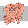
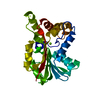
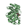
- PDB-2xtv: Structure of E.coli rhomboid protease GlpG, active site mutant, S... -
+ Open data
- Basic information
| Entry | Database: PDB / ID: 2xtv | ||||||
|---|---|---|---|---|---|---|---|
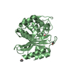
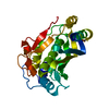
| Title | Structure of E.coli rhomboid protease GlpG, active site mutant, S201T, orthorhombic crystal form | ||||||
 Components Components | RHOMBOID PROTEASE GLPG | ||||||
 Keywords Keywords | HYDROLASE / MEMBRANE PROTEIN | ||||||
| Function / homology |  Function and homology information Function and homology informationrhomboid protease / endopeptidase activity / serine-type endopeptidase activity / proteolysis / identical protein binding / plasma membrane Similarity search - Function | ||||||
| Biological species |  | ||||||
| Method |  X-RAY DIFFRACTION / X-RAY DIFFRACTION /  SYNCHROTRON / SYNCHROTRON /  MOLECULAR REPLACEMENT / Resolution: 1.7 Å MOLECULAR REPLACEMENT / Resolution: 1.7 Å | ||||||
 Authors Authors | Vinothkumar, K.R. | ||||||
 Citation Citation |  Journal: J. Mol. Biol. / Year: 2011 Journal: J. Mol. Biol. / Year: 2011Title: Structure of rhomboid protease in a lipid environment. Authors: Vinothkumar, K.R. | ||||||
| History |
|
- Structure visualization
| Structure viewer | Molecule:  Molmil Molmil Jmol/JSmol Jmol/JSmol |
|---|
- Downloads & links
- Download
| PDBx/mmCIF format |  2xtv.cif.gz 2xtv.cif.gz | 62 KB | Display |  PDBx/mmCIF format PDBx/mmCIF format |
|---|---|---|---|---|
| PDB format |  pdb2xtv.ent.gz pdb2xtv.ent.gz | 42.8 KB | Display |  PDB format PDB format |
| PDBx/mmJSON format |  2xtv.json.gz 2xtv.json.gz | Tree view |  PDBx/mmJSON format PDBx/mmJSON format | |
| Others |  Other downloads Other downloads |
-Validation report
| Summary document |  2xtv_validation.pdf.gz 2xtv_validation.pdf.gz | 2.2 MB | Display |  wwPDB validaton report wwPDB validaton report |
|---|---|---|---|---|
| Full document |  2xtv_full_validation.pdf.gz 2xtv_full_validation.pdf.gz | 2.2 MB | Display | |
| Data in XML |  2xtv_validation.xml.gz 2xtv_validation.xml.gz | 12.4 KB | Display | |
| Data in CIF |  2xtv_validation.cif.gz 2xtv_validation.cif.gz | 15.9 KB | Display | |
| Arichive directory |  https://data.pdbj.org/pub/pdb/validation_reports/xt/2xtv https://data.pdbj.org/pub/pdb/validation_reports/xt/2xtv ftp://data.pdbj.org/pub/pdb/validation_reports/xt/2xtv ftp://data.pdbj.org/pub/pdb/validation_reports/xt/2xtv | HTTPS FTP |
-Related structure data
| Related structure data |  2xtuC 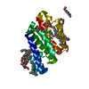 2xovS S: Starting model for refinement C: citing same article ( |
|---|---|
| Similar structure data |
- Links
Links
- Assembly
| Deposited unit | 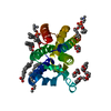
| ||||||||
|---|---|---|---|---|---|---|---|---|---|
| 1 |
| ||||||||
| Unit cell |
|
- Components
| #1: Protein | Mass: 20256.033 Da / Num. of mol.: 1 / Fragment: CORE TM DOMAIN, RESIDUES 93-272 / Mutation: YES Source method: isolated from a genetically manipulated source Source: (gene. exp.)   | ||||||
|---|---|---|---|---|---|---|---|
| #2: Chemical | ChemComp-MC3 / #3: Water | ChemComp-HOH / | Compound details | ENGINEERED | Nonpolymer details | IN THE PRESENT MODEL LIPIDS (MC3) MOLECULES 504, 505, 508, 511, 513 AND 514 COULD ALSO REPRESENT ...IN THE PRESENT MODEL LIPIDS (MC3) MOLECULES 504, 505, 508, 511, 513 AND 514 COULD ALSO REPRESENT DETERGENT NONYL GLUCOSIDE USED FOR PURIFICATI | |
-Experimental details
-Experiment
| Experiment | Method:  X-RAY DIFFRACTION / Number of used crystals: 1 X-RAY DIFFRACTION / Number of used crystals: 1 |
|---|
- Sample preparation
| Crystal | Density Matthews: 2.41 Å3/Da / Density % sol: 48.94 % / Description: NONE |
|---|---|
| Crystal grow | Temperature: 298 K / Method: vapor diffusion, hanging drop / pH: 7 Details: 1.5M NACL, 0.1M BIS-TRIS, PH7, 2% DMPC/CHAPSO, (ADDED ONLY TO THE PROTEIN), 298K, pH 7.0 |
-Data collection
| Diffraction | Mean temperature: 100 K |
|---|---|
| Diffraction source | Source:  SYNCHROTRON / Site: SYNCHROTRON / Site:  Diamond Diamond  / Beamline: I03 / Wavelength: 0.9763 / Beamline: I03 / Wavelength: 0.9763 |
| Detector | Type: ADSC CCD / Detector: CCD / Date: Apr 29, 2010 |
| Radiation | Protocol: SINGLE WAVELENGTH / Monochromatic (M) / Laue (L): M / Scattering type: x-ray |
| Radiation wavelength | Wavelength: 0.9763 Å / Relative weight: 1 |
| Reflection | Resolution: 1.7→45.54 Å / Num. obs: 22469 / % possible obs: 95.8 % / Observed criterion σ(I): 0 / Redundancy: 3.3 % / Biso Wilson estimate: 20.3 Å2 / Rmerge(I) obs: 0.05 / Net I/σ(I): 13.3 |
| Reflection shell | Resolution: 1.7→1.79 Å / Redundancy: 3.1 % / Rmerge(I) obs: 0.31 / Mean I/σ(I) obs: 3.2 / % possible all: 86.9 |
- Processing
| Software |
| |||||||||||||||||||||||||||||||||||||||||||||||||||||||||||||||
|---|---|---|---|---|---|---|---|---|---|---|---|---|---|---|---|---|---|---|---|---|---|---|---|---|---|---|---|---|---|---|---|---|---|---|---|---|---|---|---|---|---|---|---|---|---|---|---|---|---|---|---|---|---|---|---|---|---|---|---|---|---|---|---|---|
| Refinement | Method to determine structure:  MOLECULAR REPLACEMENT MOLECULAR REPLACEMENTStarting model: PDB ENTRY 2XOV Resolution: 1.7→28.009 Å / SU ML: 0.19 / σ(F): 0 / Phase error: 18.38 / Stereochemistry target values: ML Details: RESIDUES 246 AND 247 ARE DISORDERED. HENCE THERE IS A GAP BETWEEN 245 AND 248.THERE IS A DENSITY ABOVE THE ACTIVE SITE WHICH HAS NOT BEEN MODELLED, AS NONE OF COMPONENTS OF CRYSTALLISATION ...Details: RESIDUES 246 AND 247 ARE DISORDERED. HENCE THERE IS A GAP BETWEEN 245 AND 248.THERE IS A DENSITY ABOVE THE ACTIVE SITE WHICH HAS NOT BEEN MODELLED, AS NONE OF COMPONENTS OF CRYSTALLISATION CAN EXPLAIN THE DENSITY.
| |||||||||||||||||||||||||||||||||||||||||||||||||||||||||||||||
| Solvent computation | Shrinkage radii: 0.9 Å / VDW probe radii: 1.11 Å / Solvent model: FLAT BULK SOLVENT MODEL / Bsol: 66.136 Å2 / ksol: 0.412 e/Å3 | |||||||||||||||||||||||||||||||||||||||||||||||||||||||||||||||
| Displacement parameters | Biso mean: 24.1 Å2
| |||||||||||||||||||||||||||||||||||||||||||||||||||||||||||||||
| Refinement step | Cycle: LAST / Resolution: 1.7→28.009 Å
| |||||||||||||||||||||||||||||||||||||||||||||||||||||||||||||||
| Refine LS restraints |
| |||||||||||||||||||||||||||||||||||||||||||||||||||||||||||||||
| LS refinement shell |
|
 Movie
Movie Controller





 PDBj
PDBj