Protocol: SINGLE WAVELENGTH / Monochromatic (M) / Laue (L): M / Scattering type: x-ray
Radiation wavelength
Wavelength: 0.978 Å / Relative weight: 1
Reflection
Resolution: 1.9→20 Å / Num. obs: 20683 / % possible obs: 99.7 % / Observed criterion σ(I): 1 / Redundancy: 5.2 % / Biso Wilson estimate: 36 Å2 / Rmerge(I) obs: 0.09 / Net I/σ(I): 10.9
Reflection shell
Resolution: 1.9→2 Å / Redundancy: 5.4 % / Rmerge(I) obs: 0.66 / Mean I/σ(I) obs: 2.3 / % possible all: 98.3
-
Processing
Software
Name
Version
Classification
XDS
datareduction
SCALA
datascaling
PHENIX.HYSS
phasing
PHASER
phasing
REFMAC
5.6.0081
refinement
Refinement
Method to determine structure: SAD Starting model: NONE Resolution: 1.9→58.32 Å / Cor.coef. Fo:Fc: 0.954 / Cor.coef. Fo:Fc free: 0.951 / SU B: 4.741 / SU ML: 0.073 / Cross valid method: THROUGHOUT / ESU R: 0.115 / ESU R Free: 0.109 / Stereochemistry target values: MAXIMUM LIKELIHOOD Details: HYDROGENS HAVE BEEN ADDED IN THE RIDING POSITIONS. ATOM RECORD CONTAINS SUM OF TLS AND RESIDUAL B FACTORS. ANISOU RECORD CONTAINS SUM OF TLS AND RESIDUAL U FACTORS.
Rfactor
Num. reflection
% reflection
Selection details
Rfree
0.20038
1055
5.1 %
RANDOM
Rwork
0.1742
-
-
-
obs
0.17553
19541
99.58 %
-
Solvent computation
Ion probe radii: 0.8 Å / Shrinkage radii: 0.8 Å / VDW probe radii: 1.2 Å / Solvent model: BABINET MODEL WITH MASK
 Movie
Movie Controller

 Yorodumi
 Open data
 Basic information
 Components
 Keywords
 Function and homology information
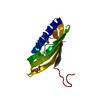
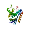
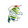
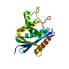
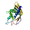
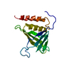
 LEISHMANIA TARENTOLAE (eukaryote)
 X-RAY DIFFRACTION /
  Authors
 Citation
 Structure visualization
 Downloads & links
 Other downloads
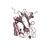


 PDBj
PDBj Assembly


 Mass: 62.005 Da / Num. of mol.: 1 / Source method: obtained synthetically / Formula: NO3
 Mass: 92.094 Da / Num. of mol.: 1 / Source method: obtained synthetically / Formula: C3H8O3
 Mass: 18.015 Da / Num. of mol.: 164 / Source method: isolated from a natural source / Formula: H2O
 Sample preparation
 / Beamline: X06SA / Wavelength: 0.978
  Processing