 Movie
Movie Controller

[English] 日本語
 Yorodumi
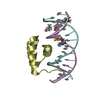
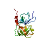
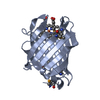
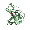
- PDB-2w56: Structure of the hypothetical protein VC0508 from Vibrio cholerae... -
+ Open data

- Basic information
| Entry | Database: PDB / ID: 2w56 | ||||||
|---|---|---|---|---|---|---|---|
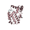
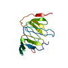
| Title | Structure of the hypothetical protein VC0508 from Vibrio cholerae VSP- II pathogenicity island | ||||||
 Components Components | (VC0508) x 2 | ||||||
 Keywords Keywords | UNKNOWN FUNCTION | ||||||
| Function / homology | Protein of unknown function DUF2787 / Protein of unknown function DUF2787 / Protein of unknown function (DUF2787) / Nuclear Transport Factor 2; Chain: A, / Roll / Alpha Beta / DUF2787 domain-containing protein Function and homology information Function and homology information | ||||||
| Biological species |   VIBRIO CHOLERAE (bacteria) VIBRIO CHOLERAE (bacteria) | ||||||
| Method |  X-RAY DIFFRACTION / MOLECULAR REPLACEMENT / Resolution: 1.9 Å X-RAY DIFFRACTION / MOLECULAR REPLACEMENT / Resolution: 1.9 Å | ||||||
 Authors Authors | Sheikh, M.A. / Taylor, G.L. | ||||||
 Citation Citation | Journal: To be Published Title: Structure of the Hypothetical Protein Vc0508 from Vibrio Cholerae Vsp-II Pathogenicity Island Authors: Sheikh, M.A. / Taylor, G.L. | ||||||
| History |
|
- Structure visualization
| Structure viewer | Molecule: MolmilJmol/JSmol |
|---|
- Downloads & links
-Download
| PDBx/mmCIF format | 2w56.cif.gz | 71.9 KB | Display | PDBx/mmCIF format |
|---|---|---|---|---|
| PDB format | pdb2w56.ent.gz | 53.3 KB | Display | PDB format |
| PDBx/mmJSON format | 2w56.json.gz | Tree view | PDBx/mmJSON format | |
| Others |  Other downloads Other downloads |
-Validation report
| Arichive directory | https://data.pdbj.org/pub/pdb/validation_reports/w5/2w56ftp://data.pdbj.org/pub/pdb/validation_reports/w5/2w56 | HTTPS FTP |
|---|
-Related structure data
| Related structure data |  2v1lS S: Starting model for refinement |
|---|---|
| Similar structure data |


-Links
 PDBj
PDBj- Assembly
| Deposited unit | 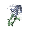
| ||||||||
|---|---|---|---|---|---|---|---|---|---|
| 1 | 
| ||||||||
| 2 | 
| ||||||||
| Unit cell |
|
-Components
| #1: Protein | Mass: 17022.184 Da / Num. of mol.: 1 Source method: isolated from a genetically manipulated source Source: (gene. exp.) VIBRIO CHOLERAE (bacteria) / Strain: N16961 / Production host: |
|---|---|
| #2: Protein | Mass: 17022.117 Da / Num. of mol.: 1 Source method: isolated from a genetically manipulated source Source: (gene. exp.) VIBRIO CHOLERAE (bacteria) / Strain: N16961 / Production host: |
| #3: Water | ChemComp-HOH /  Mass: 18.015 Da / Num. of mol.: 194 / Source method: isolated from a natural source / Formula: H2O Mass: 18.015 Da / Num. of mol.: 194 / Source method: isolated from a natural source / Formula: H2O |
-Experimental details
-Experiment
| Experiment | Method: X-RAY DIFFRACTION |
|---|
- Sample preparation
| Crystal | Density Matthews: 2.05 Å3/Da / Density % sol: 40 % / Description: NONE |
|---|---|
| Crystal grow | pH: 5.5 / Details: PH 5.5 |
-Data collection
| Diffraction | Mean temperature: 100 K |
|---|---|
| Diffraction source | Source: ROTATING ANODE / Type: RIGAKU MICROMAX-007 HF / Wavelength: 1.5418 |
| Detector | Type: RIGAKU SATURN 944 / Detector: CCD |
| Radiation | Protocol: SINGLE WAVELENGTH / Monochromatic (M) / Laue (L): M / Scattering type: x-ray |
| Radiation wavelength | Wavelength: 1.5418 Å / Relative weight: 1 |
| Reflection | Resolution: 1.9→20 Å / Num. obs: 21026 / % possible obs: 97 % / Observed criterion σ(I): 0 / Redundancy: 6.5 % / Rmerge(I) obs: 0.03 / Net I/σ(I): 62 |
| Reflection shell | Resolution: 1.9→1.97 Å / Redundancy: 6.2 % / Rmerge(I) obs: 0.03 / Mean I/σ(I) obs: 5.6 / % possible all: 96 |
- Processing
| Software |
| ||||||||||||||||||||||||||||||||||||||||||||||||||||||||||||||||||||||||||||||||||||||||||||||||||||||||||||||||||||||||||||||||||||||||||||||||||||||||||||||||||||||||||||||||||||||
|---|---|---|---|---|---|---|---|---|---|---|---|---|---|---|---|---|---|---|---|---|---|---|---|---|---|---|---|---|---|---|---|---|---|---|---|---|---|---|---|---|---|---|---|---|---|---|---|---|---|---|---|---|---|---|---|---|---|---|---|---|---|---|---|---|---|---|---|---|---|---|---|---|---|---|---|---|---|---|---|---|---|---|---|---|---|---|---|---|---|---|---|---|---|---|---|---|---|---|---|---|---|---|---|---|---|---|---|---|---|---|---|---|---|---|---|---|---|---|---|---|---|---|---|---|---|---|---|---|---|---|---|---|---|---|---|---|---|---|---|---|---|---|---|---|---|---|---|---|---|---|---|---|---|---|---|---|---|---|---|---|---|---|---|---|---|---|---|---|---|---|---|---|---|---|---|---|---|---|---|---|---|---|---|
| Refinement | Method to determine structure: MOLECULAR REPLACEMENT Starting model: PDB ENTRY 2V1L Resolution: 1.9→48.06 Å / Cor.coef. Fo:Fc: 0.945 / Cor.coef. Fo:Fc free: 0.916 / SU B: 9.055 / SU ML: 0.136 / Cross valid method: THROUGHOUT / ESU R: 0.216 / ESU R Free: 0.189 / Stereochemistry target values: MAXIMUM LIKELIHOOD / Details: HYDROGENS HAVE BEEN ADDED IN THE RIDING POSITIONS.
| ||||||||||||||||||||||||||||||||||||||||||||||||||||||||||||||||||||||||||||||||||||||||||||||||||||||||||||||||||||||||||||||||||||||||||||||||||||||||||||||||||||||||||||||||||||||
| Solvent computation | Ion probe radii: 0.8 Å / Shrinkage radii: 0.8 Å / VDW probe radii: 1.2 Å / Solvent model: BABINET MODEL WITH MASK | ||||||||||||||||||||||||||||||||||||||||||||||||||||||||||||||||||||||||||||||||||||||||||||||||||||||||||||||||||||||||||||||||||||||||||||||||||||||||||||||||||||||||||||||||||||||
| Displacement parameters | Biso mean: 36.22 Å2
| ||||||||||||||||||||||||||||||||||||||||||||||||||||||||||||||||||||||||||||||||||||||||||||||||||||||||||||||||||||||||||||||||||||||||||||||||||||||||||||||||||||||||||||||||||||||
| Refinement step | Cycle: LAST / Resolution: 1.9→48.06 Å
| ||||||||||||||||||||||||||||||||||||||||||||||||||||||||||||||||||||||||||||||||||||||||||||||||||||||||||||||||||||||||||||||||||||||||||||||||||||||||||||||||||||||||||||||||||||||
| Refine LS restraints |
|