Mass: 18.015 Da / Num. of mol.: 114 / Source method: isolated from a natural source / Formula: H2O
Nonpolymer details
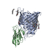
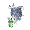
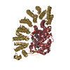
GLUTATHIONE (GSH): ATTACHED TO CYS147
-
Experimental details
-
Experiment
Experiment
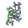
Method: X-RAY DIFFRACTION / Number of used crystals: 1
-
Sample preparation
Crystal
Density Matthews: 1.95 Å3/Da / Density % sol: 37 % / Description: NONE
Crystal grow
Method: vapor diffusion, hanging drop Details: TO OBTAIN DIFFRACTION QUALITY CRYSTALS, HANGING DROPS OF 2 UL OF PROTEIN (6 MG/ML) AND 1 UL OF MOTHER LIQUOR (0.1 M MMT BUFFER, PH 8, 12-14% PEG 6000) WERE ALLOWED TO EQUILIBRATE AGAINST THE ...Details: TO OBTAIN DIFFRACTION QUALITY CRYSTALS, HANGING DROPS OF 2 UL OF PROTEIN (6 MG/ML) AND 1 UL OF MOTHER LIQUOR (0.1 M MMT BUFFER, PH 8, 12-14% PEG 6000) WERE ALLOWED TO EQUILIBRATE AGAINST THE MOTHER LIQUOR FOR 18-24 HOURS BEFORE THE DROPS WERE STREAK SEEDED FROM EARLIER CRYSTALS.
-
Data collection
Diffraction
Mean temperature: 100 K
Diffraction source
Source: SYNCHROTRON / Site: MAX II / Beamline: I911-2 / Wavelength: 1.04
Detector
Type: MARRESEARCH / Detector: CCD / Details: MULTILAYER MIRROR, CURVED TO FOCUS IN THE VERTICAL
Radiation
Monochromator: BENT SI (111) CRYSTAL, HORIZONTALLY FOCUSING / Protocol: SINGLE WAVELENGTH / Monochromatic (M) / Laue (L): M / Scattering type: x-ray
Radiation wavelength
Wavelength: 1.04 Å / Relative weight: 1
Reflection
Resolution: 2.7→26 Å / Num. obs: 30000 / % possible obs: 95.3 % / Observed criterion σ(I): 0 / Redundancy: 4.1 % / Biso Wilson estimate: 43.62 Å2 / Rmerge(I) obs: 0.09 / Net I/σ(I): 7.4
Reflection shell
Resolution: 2.7→2.75 Å / Redundancy: 3.6 % / Rmerge(I) obs: 0.47 / Mean I/σ(I) obs: 1.6 / % possible all: 68.8
In the structure databanks used in Yorodumi, some data are registered as the other names, "COVID-19 virus" and "2019-nCoV". Here are the details of the virus and the list of structure data.
Jan 31, 2019. EMDB accession codes are about to change! (news from PDBe EMDB page)
EMDB accession codes are about to change! (news from PDBe EMDB page)
The allocation of 4 digits for EMDB accession codes will soon come to an end. Whilst these codes will remain in use, new EMDB accession codes will include an additional digit and will expand incrementally as the available range of codes is exhausted. The current 4-digit format prefixed with “EMD-” (i.e. EMD-XXXX) will advance to a 5-digit format (i.e. EMD-XXXXX), and so on. It is currently estimated that the 4-digit codes will be depleted around Spring 2019, at which point the 5-digit format will come into force.
The EM Navigator/Yorodumi systems omit the EMD- prefix.
Related info.:Q: What is EMD? / ID/Accession-code notation in Yorodumi/EM Navigator
Yorodumi is a browser for structure data from EMDB, PDB, SASBDB, etc.
This page is also the successor to EM Navigator detail page, and also detail information page/front-end page for Omokage search.
The word "yorodu" (or yorozu) is an old Japanese word meaning "ten thousand". "mi" (miru) is to see.
Related info.:EMDB / PDB / SASBDB / Comparison of 3 databanks / Yorodumi Search / Aug 31, 2016. New EM Navigator & Yorodumi / Yorodumi Papers / Jmol/JSmol / Function and homology information / Changes in new EM Navigator and Yorodumi
 Movie
Movie Controller

 Yorodumi
 Open data
 Basic information
 Components
 Keywords
 Function and homology information
 X-RAY DIFFRACTION /
  Authors
 Citation
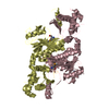
 Structure visualization
 Downloads & links
 Other downloads
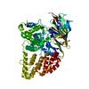


 PDBj
PDBj


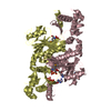
 Assembly
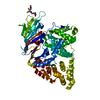


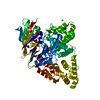
 Mass: 92.094 Da / Num. of mol.: 1 / Source method: obtained synthetically / Formula: C3H8O3
 Mass: 65.409 Da / Num. of mol.: 1 / Source method: obtained synthetically / Formula: Zn
 Mass: 307.323 Da / Num. of mol.: 1 / Source method: obtained synthetically / Formula: C10H17N3O6S
 Mass: 18.015 Da / Num. of mol.: 114 / Source method: isolated from a natural source / Formula: H2O
 Sample preparation
 / Beamline: I911-2 / Wavelength: 1.04
  Processing