 Movie
Movie Controller

[English] 日本語
 Yorodumi
- PDB-2xhn: Rhamnogalacturonan lyase from Aspergillus aculeatus K150A active ... -
+ Open data

- Basic information
| Entry | Database: PDB / ID: 2xhn | |||||||||
|---|---|---|---|---|---|---|---|---|---|---|
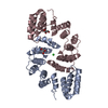
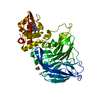
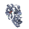
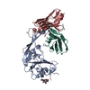
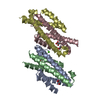
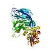
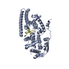
| Title | Rhamnogalacturonan lyase from Aspergillus aculeatus K150A active site mutant | |||||||||
 Components Components | RHAMNOGALACTURONASE B | |||||||||
 Keywords Keywords | LYASE / CARBOHYDRATE ACTIVE ENZYME / PECTIN / DEGRADATION / POLYSACCHARIDE LYASE FAMILY 4 | |||||||||
| Function / homology |  Function and homology information Function and homology informationrhamnogalacturonan endolyase / rhamnogalacturonan endolyase activity / pectin catabolic process / cell wall organization / carbohydrate binding / extracellular region Similarity search - Function | |||||||||
| Biological species |  | |||||||||
| Method |  X-RAY DIFFRACTION / SYNCHROTRON / MOLECULAR REPLACEMENT / Resolution: 1.52 Å X-RAY DIFFRACTION / SYNCHROTRON / MOLECULAR REPLACEMENT / Resolution: 1.52 Å | |||||||||
 Authors Authors | Jensen, M.H. / Otten, H. / Christensen, U. / Borchert, T.V. / Christensen, L.L.H. / Larsen, S. / Lo Leggio, L. | |||||||||
 Citation Citation | Journal: J.Mol.Biol. / Year: 2010 Title: Structural and Biochemical Studies Elucidate the Mechanism of Rhamnogalacturonan Lyase from Aspergillus Aculeatus. Authors: Jensen, M.H. / Otten, H. / Christensen, U. / Borchert, T.V. / Christensen, L.L.H. / Larsen, S. / Leggio, L.L. | |||||||||
| History |
|
- Structure visualization
| Structure viewer | Molecule: MolmilJmol/JSmol |
|---|
- Downloads & links
-Download
| PDBx/mmCIF format | 2xhn.cif.gz | 593.8 KB | Display | PDBx/mmCIF format |
|---|---|---|---|---|
| PDB format | pdb2xhn.ent.gz | 494.7 KB | Display | PDB format |
| PDBx/mmJSON format | 2xhn.json.gz | Tree view | PDBx/mmJSON format | |
| Others |  Other downloads Other downloads |
-Validation report
| Arichive directory | https://data.pdbj.org/pub/pdb/validation_reports/xh/2xhnftp://data.pdbj.org/pub/pdb/validation_reports/xh/2xhn | HTTPS FTP |
|---|
-Related structure data
| Related structure data | 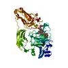 3njvC 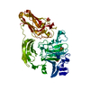 3njxC 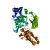 1nkgS S: Starting model for refinement C: citing same article ( |
|---|---|
| Similar structure data |


-Links
 PDBj
PDBj


- Assembly
| Deposited unit | 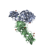
| ||||||||||||
|---|---|---|---|---|---|---|---|---|---|---|---|---|---|
| 1 | 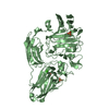
| ||||||||||||
| 2 | 
| ||||||||||||
| Unit cell |
| ||||||||||||
| Components on special symmetry positions |
|
-Components
| #1: Protein | Mass: 54185.422 Da / Num. of mol.: 2 / Mutation: YES Source method: isolated from a genetically manipulated source Source: (gene. exp.) #2: Chemical |   Mass: 40.078 Da / Num. of mol.: 2 / Source method: obtained synthetically / Formula: Ca Mass: 40.078 Da / Num. of mol.: 2 / Source method: obtained synthetically / Formula: Ca#3: Chemical | ChemComp-SO4 /   Mass: 96.063 Da / Num. of mol.: 8 / Source method: obtained synthetically / Formula: SO4 Mass: 96.063 Da / Num. of mol.: 8 / Source method: obtained synthetically / Formula: SO4#4: Chemical | ChemComp-EDO / |   Mass: 62.068 Da / Num. of mol.: 1 / Source method: obtained synthetically / Formula: C2H6O2 Mass: 62.068 Da / Num. of mol.: 1 / Source method: obtained synthetically / Formula: C2H6O2#5: Water | ChemComp-HOH / |  Mass: 18.015 Da / Num. of mol.: 1880 / Source method: isolated from a natural source / Formula: H2O Mass: 18.015 Da / Num. of mol.: 1880 / Source method: isolated from a natural source / Formula: H2OCompound details | ENGINEERED | Has protein modification | Y | |
|---|
-Experimental details
-Experiment
| Experiment | Method: X-RAY DIFFRACTION / Number of used crystals: 1 |
|---|
- Sample preparation
| Crystal | Density Matthews: 2.3 Å3/Da / Density % sol: 47 % / Description: NONE |
|---|---|
| Crystal grow | Method: vapor diffusion, hanging drop Details: 20% PEG 4000, 0.1 AMMONIUM SULFATE, HANGING DROP VAPOR DIFFUSION |
-Data collection
| Diffraction | Mean temperature: 100 K |
|---|---|
| Diffraction source | Source: SYNCHROTRON / Site: MAX II  / Beamline: I911-5 / Wavelength: 0.97 / Beamline: I911-5 / Wavelength: 0.97 |
| Detector | Type: MARRESEARCH / Detector: CCD / Date: Feb 16, 2006 / Details: MULTILAYER MIRROR, CURVED TO FOCUS IN THE VERTICAL |
| Radiation | Monochromator: BENT SI (220) CRYSTAL / Protocol: SINGLE WAVELENGTH / Monochromatic (M) / Laue (L): M / Scattering type: x-ray |
| Radiation wavelength | Wavelength: 0.97 Å / Relative weight: 1 |
| Reflection | Resolution: 1.52→20 Å / Num. obs: 142062 / % possible obs: 91.6 % / Observed criterion σ(I): -3 / Redundancy: 4.9 % / Biso Wilson estimate: 10.01 Å2 / Rmerge(I) obs: 0.06 / Net I/σ(I): 24 |
| Reflection shell | Resolution: 1.52→1.57 Å / Redundancy: 4.6 % / Rmerge(I) obs: 0.15 / Mean I/σ(I) obs: 9.7 / % possible all: 86 |
- Processing
| Software |
| |||||||||||||||||||||||||||||||||||||||||||||||||||||||||||||||||||||||||||||||||||||||||||||||||||||||||||||||||||||||||||||||||||||||||||||||||||||||||||||||||||||||||||||||||||||||||||||||||||||||||||||||||||||||||
|---|---|---|---|---|---|---|---|---|---|---|---|---|---|---|---|---|---|---|---|---|---|---|---|---|---|---|---|---|---|---|---|---|---|---|---|---|---|---|---|---|---|---|---|---|---|---|---|---|---|---|---|---|---|---|---|---|---|---|---|---|---|---|---|---|---|---|---|---|---|---|---|---|---|---|---|---|---|---|---|---|---|---|---|---|---|---|---|---|---|---|---|---|---|---|---|---|---|---|---|---|---|---|---|---|---|---|---|---|---|---|---|---|---|---|---|---|---|---|---|---|---|---|---|---|---|---|---|---|---|---|---|---|---|---|---|---|---|---|---|---|---|---|---|---|---|---|---|---|---|---|---|---|---|---|---|---|---|---|---|---|---|---|---|---|---|---|---|---|---|---|---|---|---|---|---|---|---|---|---|---|---|---|---|---|---|---|---|---|---|---|---|---|---|---|---|---|---|---|---|---|---|---|---|---|---|---|---|---|---|---|---|---|---|---|---|---|---|---|
| Refinement | Method to determine structure: MOLECULAR REPLACEMENT Starting model: PDB ENTRY 1NKG Resolution: 1.52→20.007 Å / SU ML: 0.17 / σ(F): 1.34 / Phase error: 15.82 / Stereochemistry target values: ML Details: RESIDUES GLN20, ASN435 AND THR493 ARE IN DISALLOWED REGIONS OF THE RAMACHANDRAN PLOT IN BOTH CHAINS, BUT WELL SUPPORTED BY ELECTRON DENSITY AS IN THE NATIVE PDB 1NKG. THE LEU377 RESIDUE IS ...Details: RESIDUES GLN20, ASN435 AND THR493 ARE IN DISALLOWED REGIONS OF THE RAMACHANDRAN PLOT IN BOTH CHAINS, BUT WELL SUPPORTED BY ELECTRON DENSITY AS IN THE NATIVE PDB 1NKG. THE LEU377 RESIDUE IS DISORDERED IN BOTH CHAINS.
| |||||||||||||||||||||||||||||||||||||||||||||||||||||||||||||||||||||||||||||||||||||||||||||||||||||||||||||||||||||||||||||||||||||||||||||||||||||||||||||||||||||||||||||||||||||||||||||||||||||||||||||||||||||||||
| Solvent computation | Shrinkage radii: 0.9 Å / VDW probe radii: 1.11 Å / Solvent model: FLAT BULK SOLVENT MODEL / Bsol: 47.768 Å2 / ksol: 0.409 e/Å3 | |||||||||||||||||||||||||||||||||||||||||||||||||||||||||||||||||||||||||||||||||||||||||||||||||||||||||||||||||||||||||||||||||||||||||||||||||||||||||||||||||||||||||||||||||||||||||||||||||||||||||||||||||||||||||
| Displacement parameters |
| |||||||||||||||||||||||||||||||||||||||||||||||||||||||||||||||||||||||||||||||||||||||||||||||||||||||||||||||||||||||||||||||||||||||||||||||||||||||||||||||||||||||||||||||||||||||||||||||||||||||||||||||||||||||||
| Refinement step | Cycle: LAST / Resolution: 1.52→20.007 Å
| |||||||||||||||||||||||||||||||||||||||||||||||||||||||||||||||||||||||||||||||||||||||||||||||||||||||||||||||||||||||||||||||||||||||||||||||||||||||||||||||||||||||||||||||||||||||||||||||||||||||||||||||||||||||||
| Refine LS restraints |
| |||||||||||||||||||||||||||||||||||||||||||||||||||||||||||||||||||||||||||||||||||||||||||||||||||||||||||||||||||||||||||||||||||||||||||||||||||||||||||||||||||||||||||||||||||||||||||||||||||||||||||||||||||||||||
| LS refinement shell |
|
