 Movie
Movie Controller

[English] 日本語
 Yorodumi
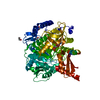
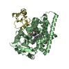
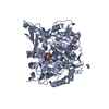
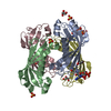
- PDB-2xfl: Induced-fit and allosteric effects upon polyene binding revealed ... -
+ Open data

- Basic information
| Entry | Database: PDB / ID: 2xfl | ||||||
|---|---|---|---|---|---|---|---|
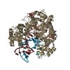
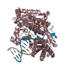
| Title | Induced-fit and allosteric effects upon polyene binding revealed by crystal structures of the Dynemicin thioesterase | ||||||
 Components Components | DYNE7 | ||||||
 Keywords Keywords | HYDROLASE / POLYKETIDE BIOSYNTHESIS / ENEDIYNE / HOT-DOG FOLD | ||||||
| Function / homology | Thioesterase-like superfamily / Hotdog Thioesterase / Thiol Ester Dehydrase; Chain A / HotDog domain superfamily / Roll / Alpha Beta / DynE7 Function and homology information Function and homology information | ||||||
| Biological species |  MICROMONOSPORA CHERSINA (bacteria) MICROMONOSPORA CHERSINA (bacteria) | ||||||
| Method |  X-RAY DIFFRACTION / SYNCHROTRON / MOLECULAR REPLACEMENT / Resolution: 2.9 Å X-RAY DIFFRACTION / SYNCHROTRON / MOLECULAR REPLACEMENT / Resolution: 2.9 Å | ||||||
 Authors Authors | Liew, C.W. / Sharff, A. / Kotaka, M. / Kong, R. / Sun, H. / Bricogne, G. / Liang, Z. / Lescar, J. | ||||||
 Citation Citation | Journal: J.Mol.Biol. / Year: 2010 Title: Induced-Fit Upon Ligand Binding Revealed by Crystal Structures of the Hot-Dog Fold Thioesterase in Dynemicin Biosynthesis. Authors: Liew, C.W. / Sharff, A. / Kotaka, M. / Kong, R. / Sun, H. / Qureshi, I. / Bricogne, G. / Liang, Z. / Lescar, J. #1: Journal: J.Biol.Chem. / Year: 2009Title: Structure and Catalytic Mechanism of the Thioesterase Cale7 in Enediyne Biosynthesis. Authors: Kotaka, M. / Kong, R. / Qureshi, I. / Ho, Q.S. / Sun, H. / Liew, C.W. / Goh, L.P. / Cheung, P. / Mu, Y. / Lescar, J. / Liang, Z. | ||||||
| History |
|
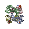
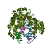
- Structure visualization
| Structure viewer | Molecule: MolmilJmol/JSmol |
|---|
- Downloads & links
-Download
| PDBx/mmCIF format | 2xfl.cif.gz | 448.3 KB | Display | PDBx/mmCIF format |
|---|---|---|---|---|
| PDB format | pdb2xfl.ent.gz | 373.9 KB | Display | PDB format |
| PDBx/mmJSON format | 2xfl.json.gz | Tree view | PDBx/mmJSON format | |
| Others |  Other downloads Other downloads |
-Validation report
| Arichive directory | https://data.pdbj.org/pub/pdb/validation_reports/xf/2xflftp://data.pdbj.org/pub/pdb/validation_reports/xf/2xfl | HTTPS FTP |
|---|
-Related structure data
| Related structure data | 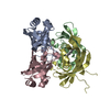 2xemC 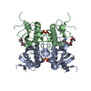 2w3xS C: citing same article ( S: Starting model for refinement |
|---|---|
| Similar structure data |

-Links
 PDBj
PDBj
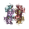
- Assembly
| Deposited unit | 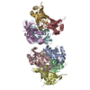
| ||||||||
|---|---|---|---|---|---|---|---|---|---|
| 1 | 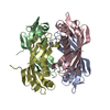
| ||||||||
| 2 | 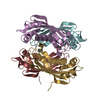
| ||||||||
| Unit cell |
|
-Components
| #1: Protein | Mass: 15880.040 Da / Num. of mol.: 8 / Fragment: RESIDUES 3-144 Source method: isolated from a genetically manipulated source Source: (gene. exp.) MICROMONOSPORA CHERSINA (bacteria) / Plasmid: PCDF-2 LIC/EK / Production host: #2: Water | ChemComp-HOH / |  Mass: 18.015 Da / Num. of mol.: 160 / Source method: isolated from a natural source / Formula: H2O Mass: 18.015 Da / Num. of mol.: 160 / Source method: isolated from a natural source / Formula: H2O |
|---|
-Experimental details
-Experiment
| Experiment | Method: X-RAY DIFFRACTION / Number of used crystals: 1 |
|---|
- Sample preparation
| Crystal | Density Matthews: 2.37 Å3/Da / Density % sol: 48 % / Description: NONE |
|---|---|
| Crystal grow | pH: 7.5 / Details: pH 7.5 |
-Data collection
| Diffraction | Mean temperature: 100 K |
|---|---|
| Diffraction source | Source: SYNCHROTRON / Site: NSRRC  / Beamline: BL13B1 / Wavelength: 1.072 / Beamline: BL13B1 / Wavelength: 1.072 |
| Detector | Type: ADSC QUANTUM 315 / Detector: CCD / Details: MIRROR |
| Radiation | Protocol: SINGLE WAVELENGTH / Monochromatic (M) / Laue (L): M / Scattering type: x-ray |
| Radiation wavelength | Wavelength: 1.072 Å / Relative weight: 1 |
| Reflection | Resolution: 2.9→40 Å / Num. obs: 27578 / % possible obs: 99 % / Observed criterion σ(I): 3 / Redundancy: 3.3 % / Biso Wilson estimate: 66.02 Å2 / Rmerge(I) obs: 0.13 / Net I/σ(I): 6.7 |
| Reflection shell | Resolution: 2.9→9.2 Å / Redundancy: 3.3 % / Rmerge(I) obs: 0.46 / Mean I/σ(I) obs: 2.3 / % possible all: 98 |
- Processing
| Software |
| |||||||||||||||||||||||||||||||||||||||||||||||||||||||||||||||||||||||||||||||||||||||||||||||||||||||||||||||||||||||||||||||||||||||||||||||||||||||||||||||||||||||||||||||||||||||||||||||||||||||||||||||||||||||||||||||||
|---|---|---|---|---|---|---|---|---|---|---|---|---|---|---|---|---|---|---|---|---|---|---|---|---|---|---|---|---|---|---|---|---|---|---|---|---|---|---|---|---|---|---|---|---|---|---|---|---|---|---|---|---|---|---|---|---|---|---|---|---|---|---|---|---|---|---|---|---|---|---|---|---|---|---|---|---|---|---|---|---|---|---|---|---|---|---|---|---|---|---|---|---|---|---|---|---|---|---|---|---|---|---|---|---|---|---|---|---|---|---|---|---|---|---|---|---|---|---|---|---|---|---|---|---|---|---|---|---|---|---|---|---|---|---|---|---|---|---|---|---|---|---|---|---|---|---|---|---|---|---|---|---|---|---|---|---|---|---|---|---|---|---|---|---|---|---|---|---|---|---|---|---|---|---|---|---|---|---|---|---|---|---|---|---|---|---|---|---|---|---|---|---|---|---|---|---|---|---|---|---|---|---|---|---|---|---|---|---|---|---|---|---|---|---|---|---|---|---|---|---|---|---|---|---|---|---|
| Refinement | Method to determine structure: MOLECULAR REPLACEMENT Starting model: PDB ENTRY 2W3X Resolution: 2.9→24.28 Å / Cor.coef. Fo:Fc: 0.8857 / Cor.coef. Fo:Fc free: 0.8476 / Cross valid method: THROUGHOUT / σ(F): 0 / SU Rfree Blow DPI: 0.392 Details: IDEAL-DIST CONTACT TERM CONTACT SETUP. ALL ATOMS HAVE CCP4 ATOM TYPE FROM LIBRARY
| |||||||||||||||||||||||||||||||||||||||||||||||||||||||||||||||||||||||||||||||||||||||||||||||||||||||||||||||||||||||||||||||||||||||||||||||||||||||||||||||||||||||||||||||||||||||||||||||||||||||||||||||||||||||||||||||||
| Displacement parameters | Biso mean: 58.05 Å2
| |||||||||||||||||||||||||||||||||||||||||||||||||||||||||||||||||||||||||||||||||||||||||||||||||||||||||||||||||||||||||||||||||||||||||||||||||||||||||||||||||||||||||||||||||||||||||||||||||||||||||||||||||||||||||||||||||
| Refine analyze | Luzzati coordinate error obs: 0.394 Å | |||||||||||||||||||||||||||||||||||||||||||||||||||||||||||||||||||||||||||||||||||||||||||||||||||||||||||||||||||||||||||||||||||||||||||||||||||||||||||||||||||||||||||||||||||||||||||||||||||||||||||||||||||||||||||||||||
| Refinement step | Cycle: LAST / Resolution: 2.9→24.28 Å
| |||||||||||||||||||||||||||||||||||||||||||||||||||||||||||||||||||||||||||||||||||||||||||||||||||||||||||||||||||||||||||||||||||||||||||||||||||||||||||||||||||||||||||||||||||||||||||||||||||||||||||||||||||||||||||||||||
| Refine LS restraints |
| |||||||||||||||||||||||||||||||||||||||||||||||||||||||||||||||||||||||||||||||||||||||||||||||||||||||||||||||||||||||||||||||||||||||||||||||||||||||||||||||||||||||||||||||||||||||||||||||||||||||||||||||||||||||||||||||||
| LS refinement shell | Resolution: 2.9→3.01 Å / Total num. of bins used: 14
| |||||||||||||||||||||||||||||||||||||||||||||||||||||||||||||||||||||||||||||||||||||||||||||||||||||||||||||||||||||||||||||||||||||||||||||||||||||||||||||||||||||||||||||||||||||||||||||||||||||||||||||||||||||||||||||||||
| Refinement TLS params. | Method: refined / Refine-ID: X-RAY DIFFRACTION
| |||||||||||||||||||||||||||||||||||||||||||||||||||||||||||||||||||||||||||||||||||||||||||||||||||||||||||||||||||||||||||||||||||||||||||||||||||||||||||||||||||||||||||||||||||||||||||||||||||||||||||||||||||||||||||||||||
| Refinement TLS group |
|
