 Movie
Movie Controller

[English] 日本語
 Yorodumi
- PDB-2x6w: Tailspike protein mutant E372Q of E.coli bacteriophage HK620 in c... -
+ Open data

- Basic information
| Entry | Database: PDB / ID: 2x6w | |||||||||
|---|---|---|---|---|---|---|---|---|---|---|
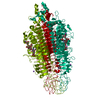
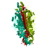
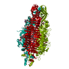
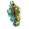
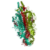
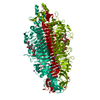
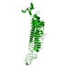
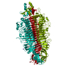
| Title | Tailspike protein mutant E372Q of E.coli bacteriophage HK620 in complex with hexasaccharide | |||||||||
 Components Components | TAIL SPIKE PROTEIN | |||||||||
 Keywords Keywords | VIRAL PROTEIN / BETA-HELIX / HYDROLASE | |||||||||
| Function / homology |  Function and homology information Function and homology informationendo-1,3-alpha-L-rhamnosidase activity / symbiont entry into host cell via disruption of host cell envelope lipopolysaccharide / virus tail, fiber / symbiont entry into host cell via disruption of host cell envelope / virion attachment to host cell / metal ion binding Similarity search - Function | |||||||||
| Biological species |  ENTEROBACTERIA PHAGE HK620 (virus) ENTEROBACTERIA PHAGE HK620 (virus) | |||||||||
| Method |  X-RAY DIFFRACTION / SYNCHROTRON / MOLECULAR REPLACEMENT / Resolution: 1.35 Å X-RAY DIFFRACTION / SYNCHROTRON / MOLECULAR REPLACEMENT / Resolution: 1.35 Å | |||||||||
 Authors Authors | Lorenzen, N.K. / Mueller, J.J. / Heinemann, U. / Seckler, R. / Barbirz, S. | |||||||||
 Citation Citation | Journal: Glycobiology / Year: 2013 Title: Single Amino Acid Exchange in Bacteriophage Hk620 Tailspike Protein Results in Thousand-Fold Increase of its Oligosaccharide Affinity. Authors: Broeker, N.K. / Gohlke, U. / Muller, J.J. / Uetrecht, C. / Heinemann, U. / Seckler, R. / Barbirz, S. #1: Journal: Mol.Microbiol. / Year: 2008Title: Crystal Structure of Escherichia Coli Phage Hk620 Tailspike: Podoviral Tailspike Endoglycosidase Modules are Evolutionarily Related. Authors: Barbirz, S. / Muller, J.J. / Uetrecht, C. / Clark, A.J. / Heinemann, U. / Seckler, R. | |||||||||
| History |
| |||||||||
| Remark 650 | HELIX DETERMINATION METHOD: AUTHOR PROVIDED. | |||||||||
| Remark 700 | SHEET DETERMINATION METHOD: AUTHOR PROVIDED. |
- Structure visualization
| Structure viewer | Molecule: MolmilJmol/JSmol |
|---|
- Downloads & links
-Download
| PDBx/mmCIF format | 2x6w.cif.gz | 278 KB | Display | PDBx/mmCIF format |
|---|---|---|---|---|
| PDB format | pdb2x6w.ent.gz | 221 KB | Display | PDB format |
| PDBx/mmJSON format | 2x6w.json.gz | Tree view | PDBx/mmJSON format | |
| Others |  Other downloads Other downloads |
-Validation report
| Arichive directory | https://data.pdbj.org/pub/pdb/validation_reports/x6/2x6wftp://data.pdbj.org/pub/pdb/validation_reports/x6/2x6w | HTTPS FTP |
|---|
-Related structure data
| Related structure data |  2x6xC  2x6yC  2x85C  4avzC  2vjiS C: citing same article ( S: Starting model for refinement |
|---|---|
| Similar structure data |

-Links
 PDBj
PDBj- Assembly
| Deposited unit | 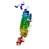
| ||||||||||||
|---|---|---|---|---|---|---|---|---|---|---|---|---|---|
| 1 | 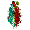
| ||||||||||||
| Unit cell |
| ||||||||||||
| Components on special symmetry positions |
|
-Components
| #1: Protein | Mass: 64704.371 Da / Num. of mol.: 1 / Fragment: RESIDUES 111-710 / Mutation: YES Source method: isolated from a genetically manipulated source Source: (gene. exp.) ENTEROBACTERIA PHAGE HK620 (virus) / Production host:  | ||||||||
|---|---|---|---|---|---|---|---|---|---|
| #2: Polysaccharide | alpha-D-glucopyranose-(1-6)-2-acetamido-2-deoxy-beta-D-glucopyranose-(1-3)-[alpha-L-rhamnopyranose- ...alpha-D-glucopyranose-(1-6)-2-acetamido-2-deoxy-beta-D-glucopyranose-(1-3)-[alpha-L-rhamnopyranose-(1-6)-alpha-D-glucopyranose-(1-4)][2-acetamido-2-deoxy-beta-D-glucopyranose-(1-3)]alpha-D-galactopyranose-(1-3)-2-acetamido-2-deoxy-alpha-D-glucopyranose Type: oligosaccharide / Mass: 1260.157 Da / Num. of mol.: 1 Source method: isolated from a genetically manipulated source | ||||||||
| #3: Chemical |   Mass: 22.990 Da / Num. of mol.: 3 / Source method: obtained synthetically / Formula: Na Mass: 22.990 Da / Num. of mol.: 3 / Source method: obtained synthetically / Formula: Na#4: Chemical | ChemComp-TRS / |   Mass: 122.143 Da / Num. of mol.: 1 / Source method: obtained synthetically / Formula: C4H12NO3 / Comment: pH buffer*YM Mass: 122.143 Da / Num. of mol.: 1 / Source method: obtained synthetically / Formula: C4H12NO3 / Comment: pH buffer*YM#5: Water | ChemComp-HOH / |  Mass: 18.015 Da / Num. of mol.: 687 / Source method: isolated from a natural source / Formula: H2O Mass: 18.015 Da / Num. of mol.: 687 / Source method: isolated from a natural source / Formula: H2OCompound details | ENGINEERED | Nonpolymer details | BOTH NAG AND NDG EXIST DUE TO MUTAROTATI | |
-Experimental details
-Experiment
| Experiment | Method: X-RAY DIFFRACTION / Number of used crystals: 1 |
|---|
- Sample preparation
| Crystal | Density Matthews: 2.15 Å3/Da / Density % sol: 42.7 % / Description: NONE |
|---|---|
| Crystal grow | Temperature: 293 K / Method: vapor diffusion, hanging drop / pH: 8.5 Details: VAPOR DIFFUSION, HANGING DROP:PROTEIN CONCENTRATION 8MG/ML. BUFFER: 40MM TRIS, PH7.8,2 MM EDTA,0.2M NACL.RERVOIR: 100MM TRIS PH8.5,3.5 MNA-FORMIATE. DROPLET 1.5:1.5 MICRO LITER. 0.3 MICRO ...Details: VAPOR DIFFUSION, HANGING DROP:PROTEIN CONCENTRATION 8MG/ML. BUFFER: 40MM TRIS, PH7.8,2 MM EDTA,0.2M NACL.RERVOIR: 100MM TRIS PH8.5,3.5 MNA-FORMIATE. DROPLET 1.5:1.5 MICRO LITER. 0.3 MICRO LITER 33MM HEXASACCHARIDE.TEMPERATURE 20 DEGREE CELSIUS. |
-Data collection
| Diffraction | Mean temperature: 100 K |
|---|---|
| Diffraction source | Source: SYNCHROTRON / Site: BESSY  / Beamline: 14.1 / Wavelength: 0.91841 / Beamline: 14.1 / Wavelength: 0.91841 |
| Detector | Type: MARRESEARCH / Detector: CCD / Date: Oct 2, 2009 / Details: GLAS MIRROR |
| Radiation | Monochromator: SI-111 CRYSTAL / Protocol: SINGLE WAVELENGTH / Monochromatic (M) / Laue (L): M / Scattering type: x-ray |
| Radiation wavelength | Wavelength: 0.91841 Å / Relative weight: 1 |
| Reflection | Resolution: 1.35→36.44 Å / Num. obs: 114674 / % possible obs: 98 % / Observed criterion σ(I): 0 / Redundancy: 3.7 % / Biso Wilson estimate: 18.3 Å2 / Rmerge(I) obs: 0.05 / Net I/σ(I): 16.2 |
| Reflection shell | Resolution: 1.35→1.39 Å / Redundancy: 2.6 % / Rmerge(I) obs: 0.34 / Mean I/σ(I) obs: 3 / % possible all: 71.5 |
- Processing
| Software |
| ||||||||||||||||||||||||||||||||||||||||||||||||||||||||||||||||||||||||||||||||||||||||||||||||||||||||||||||||||||||||||||||||||||||||||||||||||||||||||||||||||||||||||||||||||||||
|---|---|---|---|---|---|---|---|---|---|---|---|---|---|---|---|---|---|---|---|---|---|---|---|---|---|---|---|---|---|---|---|---|---|---|---|---|---|---|---|---|---|---|---|---|---|---|---|---|---|---|---|---|---|---|---|---|---|---|---|---|---|---|---|---|---|---|---|---|---|---|---|---|---|---|---|---|---|---|---|---|---|---|---|---|---|---|---|---|---|---|---|---|---|---|---|---|---|---|---|---|---|---|---|---|---|---|---|---|---|---|---|---|---|---|---|---|---|---|---|---|---|---|---|---|---|---|---|---|---|---|---|---|---|---|---|---|---|---|---|---|---|---|---|---|---|---|---|---|---|---|---|---|---|---|---|---|---|---|---|---|---|---|---|---|---|---|---|---|---|---|---|---|---|---|---|---|---|---|---|---|---|---|---|
| Refinement | Method to determine structure: MOLECULAR REPLACEMENT Starting model: PDB ENTRY 2VJI Resolution: 1.35→36.44 Å / Cor.coef. Fo:Fc: 0.98 / Cor.coef. Fo:Fc free: 0.971 / SU B: 1.565 / SU ML: 0.029 / Cross valid method: THROUGHOUT / ESU R: 0.049 / ESU R Free: 0.047 / Stereochemistry target values: MAXIMUM LIKELIHOOD Details: HYDROGENS HAVE BEEN ADDED IN THE RIDING POSITIONS. U VALUES REFINED INDIVIDUALLY.
| ||||||||||||||||||||||||||||||||||||||||||||||||||||||||||||||||||||||||||||||||||||||||||||||||||||||||||||||||||||||||||||||||||||||||||||||||||||||||||||||||||||||||||||||||||||||
| Solvent computation | Ion probe radii: 0.8 Å / Shrinkage radii: 0.8 Å / VDW probe radii: 1.4 Å / Solvent model: BABINET MODEL WITH MASK | ||||||||||||||||||||||||||||||||||||||||||||||||||||||||||||||||||||||||||||||||||||||||||||||||||||||||||||||||||||||||||||||||||||||||||||||||||||||||||||||||||||||||||||||||||||||
| Displacement parameters | Biso mean: 10.631 Å2
| ||||||||||||||||||||||||||||||||||||||||||||||||||||||||||||||||||||||||||||||||||||||||||||||||||||||||||||||||||||||||||||||||||||||||||||||||||||||||||||||||||||||||||||||||||||||
| Refinement step | Cycle: LAST / Resolution: 1.35→36.44 Å
| ||||||||||||||||||||||||||||||||||||||||||||||||||||||||||||||||||||||||||||||||||||||||||||||||||||||||||||||||||||||||||||||||||||||||||||||||||||||||||||||||||||||||||||||||||||||
| Refine LS restraints |
|