 Movie
Movie Controller

[English] 日本語
 Yorodumi
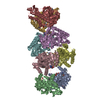
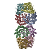
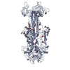
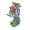
- PDB-2x0s: 3.0 A RESOLUTION CRYSTAL STRUCTURE OF GLYCOSOMAL PYRUVATE PHOSPHA... -
+ Open data

- Basic information
| Entry | Database: PDB / ID: 2x0s | |||||||||
|---|---|---|---|---|---|---|---|---|---|---|
| Title | 3.0 A RESOLUTION CRYSTAL STRUCTURE OF GLYCOSOMAL PYRUVATE PHOSPHATE DIKINASE FROM TRYPANOSOMA BRUCEI | |||||||||
 Components Components | PYRUVATE PHOSPHATE DIKINASE | |||||||||
 Keywords Keywords | TRANSFERASE / KINASE / TROPICAL PARASITE | |||||||||
| Function / homology |  Function and homology information Function and homology informationpyruvate, phosphate dikinase / pyruvate, phosphate dikinase activity / glycosome / phosphorylation / phosphate ion binding / kinase activity / ATP binding / metal ion binding Similarity search - Function | |||||||||
| Biological species |  | |||||||||
| Method |  X-RAY DIFFRACTION / SYNCHROTRON / MOLECULAR REPLACEMENT / Resolution: 2.997 Å X-RAY DIFFRACTION / SYNCHROTRON / MOLECULAR REPLACEMENT / Resolution: 2.997 Å | |||||||||
 Authors Authors | Cosenza, L.W. / Bringaud, F. / Baltz, T. / Vellieux, F.M.D. | |||||||||
 Citation Citation | Journal: J.Mol.Biol. / Year: 2001 Title: The 3.0 A Resolution Crystal Structure of Glycosomal Pyruvate Phosphate Dikinase from Trypanosoma Brucei Authors: Cosenza, L.W. / Bringaud, F. / Baltz, T. / Vellieux, F.M.D. #1: Journal: Acta Crystallogr.,Sect.D / Year: 2000 Title: Crystallization and Preliminary Crystallographic Investigation of Glycosomal Pyruvate Phosphate Dikinase from Trypanosoma Brucei Authors: Cosenza, L.W. / Bringaud, F. / Baltz, T. / Vellieux, F.M.D. #2: Journal: Proc.Natl.Acad.Sci.USA / Year: 1998 Title: Functional and Molecular Characterization of a Glycosomal Ppi-Dependent Enzyme in Trypanosomatids: Pyruvate, Phosphate Dikinase Authors: Bringaud, F. / Baltz, D. / Baltz, T. | |||||||||
| History |
| |||||||||
| Remark 700 | DETERMINATION METHOD: DSSP THE SHEETS PRESENTED AS "AF" IN EACH CHAIN ON SHEET RECORDS BELOW IS ... DETERMINATION METHOD: DSSP THE SHEETS PRESENTED AS "AF" IN EACH CHAIN ON SHEET RECORDS BELOW IS ACTUALLY AN 8-STRANDED BARREL THIS IS REPRESENTED BY A 9-STRANDED SHEET IN WHICH THE FIRST AND LAST STRANDS ARE IDENTICAL. |
- Structure visualization
| Structure viewer | Molecule: MolmilJmol/JSmol |
|---|
- Downloads & links
-Download
| PDBx/mmCIF format | 2x0s.cif.gz | 364.4 KB | Display | PDBx/mmCIF format |
|---|---|---|---|---|
| PDB format | pdb2x0s.ent.gz | 299.1 KB | Display | PDB format |
| PDBx/mmJSON format | 2x0s.json.gz | Tree view | PDBx/mmJSON format | |
| Others |  Other downloads Other downloads |
-Validation report
| Arichive directory | https://data.pdbj.org/pub/pdb/validation_reports/x0/2x0sftp://data.pdbj.org/pub/pdb/validation_reports/x0/2x0s | HTTPS FTP |
|---|
-Related structure data
| Related structure data |  1dikS S: Starting model for refinement |
|---|---|
| Similar structure data |

-Links
 PDBj
PDBj


- Assembly
| Deposited unit | 
| ||||||||
|---|---|---|---|---|---|---|---|---|---|
| 1 | 
| ||||||||
| Unit cell |
|
-Components
| #1: Protein | Mass: 100544.922 Da / Num. of mol.: 1 Source method: isolated from a genetically manipulated source Source: (gene. exp.)  |
|---|---|
| #2: Water | ChemComp-HOH /  Mass: 18.015 Da / Num. of mol.: 36 / Source method: isolated from a natural source / Formula: H2O Mass: 18.015 Da / Num. of mol.: 36 / Source method: isolated from a natural source / Formula: H2O |
-Experimental details
-Experiment
| Experiment | Method: X-RAY DIFFRACTION / Number of used crystals: 5 |
|---|
- Sample preparation
| Crystal | Density Matthews: 3.1 Å3/Da / Density % sol: 58 % / Description: NONE |
|---|---|
| Crystal grow | Method: vapor diffusion, hanging drop / pH: 8.8 Details: HANGING DROP, 2 MICROLITER PROTEIN AND 2 MICROLITER PRECIPITANT SOLUTION. PROTEIN SOLUTION. 50 MG/ML, 20 MM IMIDAZOLE PH 7.0, 100 MM NACL, 100 MM MGCL2, 20% (V/V) GLYCEROL. PRECIPITANT ...Details: HANGING DROP, 2 MICROLITER PROTEIN AND 2 MICROLITER PRECIPITANT SOLUTION. PROTEIN SOLUTION. 50 MG/ML, 20 MM IMIDAZOLE PH 7.0, 100 MM NACL, 100 MM MGCL2, 20% (V/V) GLYCEROL. PRECIPITANT SOLUTION. 0.1 M BICINE PH 8.8, 1.5 % (W/V) PEG 5000 MONOMETHYLETHER, 10 % (V/V) GLYCEROL |
-Data collection
| Diffraction | Mean temperature: 277 K |
|---|---|
| Diffraction source | Source: SYNCHROTRON / Site: LURE  / Beamline: DW32 / Wavelength: 0.964 / Beamline: DW32 / Wavelength: 0.964 |
| Detector | Type: MARRESEARCH / Detector: IMAGE PLATE / Date: Jun 15, 2000 |
| Radiation | Protocol: SINGLE WAVELENGTH / Monochromatic (M) / Laue (L): M / Scattering type: x-ray |
| Radiation wavelength | Wavelength: 0.964 Å / Relative weight: 1 |
| Reflection | Resolution: 3→24 Å / Num. obs: 24120 / % possible obs: 96 % / Observed criterion σ(I): 0 / Redundancy: 5.3 % / Biso Wilson estimate: 61 Å2 / Rmerge(I) obs: 0.09 / Net I/σ(I): 14.8 |
| Reflection shell | Resolution: 3→3.1 Å / Redundancy: 3.4 % / Rmerge(I) obs: 0.28 / Mean I/σ(I) obs: 3.1 / % possible all: 88.6 |
- Processing
| Software |
| ||||||||||||||||||||||||||||||||||||||||||||||||||||||||||||||||||||||||||||||||||||||||||||||||||||||||||||||||||||||||||||||||||||||||||||||||||||||||||||||||||||||||||||||||||||||||||||||||||||||||
|---|---|---|---|---|---|---|---|---|---|---|---|---|---|---|---|---|---|---|---|---|---|---|---|---|---|---|---|---|---|---|---|---|---|---|---|---|---|---|---|---|---|---|---|---|---|---|---|---|---|---|---|---|---|---|---|---|---|---|---|---|---|---|---|---|---|---|---|---|---|---|---|---|---|---|---|---|---|---|---|---|---|---|---|---|---|---|---|---|---|---|---|---|---|---|---|---|---|---|---|---|---|---|---|---|---|---|---|---|---|---|---|---|---|---|---|---|---|---|---|---|---|---|---|---|---|---|---|---|---|---|---|---|---|---|---|---|---|---|---|---|---|---|---|---|---|---|---|---|---|---|---|---|---|---|---|---|---|---|---|---|---|---|---|---|---|---|---|---|---|---|---|---|---|---|---|---|---|---|---|---|---|---|---|---|---|---|---|---|---|---|---|---|---|---|---|---|---|---|---|---|---|
| Refinement | Method to determine structure: MOLECULAR REPLACEMENT Starting model: PDB ENTRY 1DIK Resolution: 2.997→23.938 Å / SU ML: 0.35 / σ(F): 1.62 / Phase error: 23.59 / Stereochemistry target values: ML
| ||||||||||||||||||||||||||||||||||||||||||||||||||||||||||||||||||||||||||||||||||||||||||||||||||||||||||||||||||||||||||||||||||||||||||||||||||||||||||||||||||||||||||||||||||||||||||||||||||||||||
| Solvent computation | Shrinkage radii: 0.9 Å / VDW probe radii: 1.11 Å / Solvent model: FLAT BULK SOLVENT MODEL / Bsol: 57.101 Å2 / ksol: 0.253 e/Å3 | ||||||||||||||||||||||||||||||||||||||||||||||||||||||||||||||||||||||||||||||||||||||||||||||||||||||||||||||||||||||||||||||||||||||||||||||||||||||||||||||||||||||||||||||||||||||||||||||||||||||||
| Displacement parameters | Biso mean: 81.08 Å2
| ||||||||||||||||||||||||||||||||||||||||||||||||||||||||||||||||||||||||||||||||||||||||||||||||||||||||||||||||||||||||||||||||||||||||||||||||||||||||||||||||||||||||||||||||||||||||||||||||||||||||
| Refinement step | Cycle: LAST / Resolution: 2.997→23.938 Å
| ||||||||||||||||||||||||||||||||||||||||||||||||||||||||||||||||||||||||||||||||||||||||||||||||||||||||||||||||||||||||||||||||||||||||||||||||||||||||||||||||||||||||||||||||||||||||||||||||||||||||
| Refine LS restraints |
| ||||||||||||||||||||||||||||||||||||||||||||||||||||||||||||||||||||||||||||||||||||||||||||||||||||||||||||||||||||||||||||||||||||||||||||||||||||||||||||||||||||||||||||||||||||||||||||||||||||||||
| LS refinement shell |
| ||||||||||||||||||||||||||||||||||||||||||||||||||||||||||||||||||||||||||||||||||||||||||||||||||||||||||||||||||||||||||||||||||||||||||||||||||||||||||||||||||||||||||||||||||||||||||||||||||||||||
| Refinement TLS params. | Method: refined / Refine-ID: X-RAY DIFFRACTION
| ||||||||||||||||||||||||||||||||||||||||||||||||||||||||||||||||||||||||||||||||||||||||||||||||||||||||||||||||||||||||||||||||||||||||||||||||||||||||||||||||||||||||||||||||||||||||||||||||||||||||
| Refinement TLS group |
|
