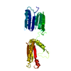
Resolution: 2.082→42.578 Å / SU ML: 0.29 / σ(F): 0.1 / Phase error: 24.26 / Stereochemistry target values: ML Details: THE LINKER RESIDUES AND RESIDUES 1061-1075 ARE DISORDERED AND NOT INCLUDED IN THE MODEL ONE ISOPEPTIDE BOND IS FORMED BETWEEN ASN1393 AND LYS1259 AND ONE ISOPEPTIDE BOND BETWEEN ASN1232 AND LYS1082
Rfactor
Num. reflection
% reflection
Rfree
0.255
1073
5.2 %
Rwork
0.1923
-
-
obs
0.1956
20786
98.16 %
Solvent computation
Shrinkage radii: 0.9 Å / VDW probe radii: 1.11 Å / Solvent model: FLAT BULK SOLVENT MODEL / Bsol: 47.965 Å2 / ksol: 0.398 e/Å3
Displacement parameters
Biso mean: 25.01 Å2
Baniso -1
Baniso -2
Baniso -3
1-
2.5025 Å2
-0 Å2
0 Å2
2-
-
-7.4428 Å2
-0 Å2
3-
-
-
5.6657 Å2
Refinement step
Cycle: LAST / Resolution: 2.082→42.578 Å
Protein
Nucleic acid
Ligand
Solvent
Total
Num. atoms
2659
0
3
138
2800
Refine LS restraints
Refine-ID
Type
Dev ideal
Number
X-RAY DIFFRACTION
f_bond_d
0.012
2711
X-RAY DIFFRACTION
f_angle_d
1.408
3677
X-RAY DIFFRACTION
f_dihedral_angle_d
18.463
977
X-RAY DIFFRACTION
f_chiral_restr
0.098
417
X-RAY DIFFRACTION
f_plane_restr
0.006
478
LS refinement shell
Resolution (Å)
Rfactor Rfree
Num. reflection Rfree
Rfactor Rwork
Num. reflection Rwork
Refine-ID
% reflection obs (%)
2.082-2.1768
0.2976
139
0.2141
2405
X-RAY DIFFRACTION
97
2.1768-2.2916
0.3062
125
0.2044
2363
X-RAY DIFFRACTION
96
2.2916-2.4351
0.2459
132
0.206
2422
X-RAY DIFFRACTION
98
2.4351-2.6231
0.3111
120
0.1947
2418
X-RAY DIFFRACTION
98
2.6231-2.887
0.247
143
0.1999
2452
X-RAY DIFFRACTION
98
2.887-3.3047
0.2451
136
0.188
2487
X-RAY DIFFRACTION
99
3.3047-4.163
0.2232
137
0.1732
2533
X-RAY DIFFRACTION
100
4.163-42.5873
0.2419
141
0.187
2633
X-RAY DIFFRACTION
99
Refinement TLS params.
Method: refined / Refine-ID: X-RAY DIFFRACTION
ID
L11 (°2)
L12 (°2)
L13 (°2)
L22 (°2)
L23 (°2)
L33 (°2)
S11 (Å °)
S12 (Å °)
S13 (Å °)
S21 (Å °)
S22 (Å °)
S23 (Å °)
S31 (Å °)
S32 (Å °)
S33 (Å °)
T11 (Å2)
T12 (Å2)
T13 (Å2)
T22 (Å2)
T23 (Å2)
T33 (Å2)
Origin x (Å)
Origin y (Å)
Origin z (Å)
1
0.17
0.2542
-0.1205
0.3551
-0.0651
0.4181
0.0032
-0.0223
0.0827
-0.0082
-0.0594
0.2022
-0.0042
-0.0778
-0
0.0865
0.0021
-0.0233
0.0892
-0.0473
0.1337
9.1943
31.5002
13.6252
2
0.5459
0.2447
0.3955
0.4743
-0.0024
0.608
-0.0093
0.0293
0.0156
-0.0031
0.0145
0.0048
0.0451
0.0253
0.0276
0.0212
0.0018
0.0112
0.0367
-0.0079
0.0218
7.24
-8.7782
-0.361
Refinement TLS group
ID
Refine-ID
Refine TLS-ID
Selection details
1
X-RAY DIFFRACTION
1
RESID1076:1253
2
X-RAY DIFFRACTION
2
RESID1254:1413
+
About Yorodumi
-
News
-
Feb 9, 2022. New format data for meta-information of EMDB entries
New format data for meta-information of EMDB entries
Version 3 of the EMDB header file is now the official format.
The previous official version 1.9 will be removed from the archive.
In the structure databanks used in Yorodumi, some data are registered as the other names, "COVID-19 virus" and "2019-nCoV". Here are the details of the virus and the list of structure data.
Jan 31, 2019. EMDB accession codes are about to change! (news from PDBe EMDB page)
EMDB accession codes are about to change! (news from PDBe EMDB page)
The allocation of 4 digits for EMDB accession codes will soon come to an end. Whilst these codes will remain in use, new EMDB accession codes will include an additional digit and will expand incrementally as the available range of codes is exhausted. The current 4-digit format prefixed with “EMD-” (i.e. EMD-XXXX) will advance to a 5-digit format (i.e. EMD-XXXXX), and so on. It is currently estimated that the 4-digit codes will be depleted around Spring 2019, at which point the 5-digit format will come into force.
The EM Navigator/Yorodumi systems omit the EMD- prefix.
Related info.:Q: What is EMD? / ID/Accession-code notation in Yorodumi/EM Navigator
Yorodumi is a browser for structure data from EMDB, PDB, SASBDB, etc.
This page is also the successor to EM Navigator detail page, and also detail information page/front-end page for Omokage search.
The word "yorodu" (or yorozu) is an old Japanese word meaning "ten thousand". "mi" (miru) is to see.
Related info.:EMDB / PDB / SASBDB / Comparison of 3 databanks / Yorodumi Search / Aug 31, 2016. New EM Navigator & Yorodumi / Yorodumi Papers / Jmol/JSmol / Function and homology information / Changes in new EM Navigator and Yorodumi
 Movie
Movie Controller

 Yorodumi
 Open data
 Basic information
 Components
 Keywords
 Function and homology information
 STREPTOCOCCUS GORDONII (bacteria)
 X-RAY DIFFRACTION /
  Authors
 Citation
 Structure visualization
 Downloads & links
 Other downloads


 PDBj
PDBj Assembly
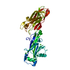

 Mass: 40.078 Da / Num. of mol.: 3 / Source method: obtained synthetically / Formula: Ca
 Mass: 18.015 Da / Num. of mol.: 138 / Source method: isolated from a natural source / Formula: H2O
 Sample preparation
 / Beamline: I911-3 / Wavelength: 0.979
  Processing