[English] 日本語
Yorodumi
- PDB-2wqs: Crystal structure of the C-terminal domain of Streptococcus gordo... -

+
Open data


ID or keywords:

Loading...

-
Basic information

Entry
Database: PDB / ID: 2wqs
TitleCrystal structure of the C-terminal domain of Streptococcus gordonii surface protein SspB
ComponentsAGGLUTININ RECEPTOR
KeywordsCELL ADHESION / CELL WALL / ANTIGEN I/II / PEPTIDOGLYCAN-ANCHOR
Function / homology
Function and homology information


extracellular region / metal ion binding
Similarity search - Function
Cell surface antigen I/II A repeat / Streptococcal surface antigen repeat / Streptococcus antigen I/II alanine-rich (Ag I/II A) repeat profile. / Glucan-binding protein C/Surface antigen I/II, V-domain / Glucan-binding protein C/Surface antigen I/II, V-domain superfamily / Glucan-binding protein C / Cross-wall-targeting lipoprotein motif / Adhesin isopeptide-forming adherence domain / Cell surface antigen, C-terminal / Antigen I/II, N-terminal ...Cell surface antigen I/II A repeat / Streptococcal surface antigen repeat / Streptococcus antigen I/II alanine-rich (Ag I/II A) repeat profile. / Glucan-binding protein C/Surface antigen I/II, V-domain / Glucan-binding protein C/Surface antigen I/II, V-domain superfamily / Glucan-binding protein C / Cross-wall-targeting lipoprotein motif / Adhesin isopeptide-forming adherence domain / Cell surface antigen, C-terminal / Antigen I/II, N-terminal / Cell surface antigen C-terminus / Cell surface antigen I/II C2 terminal domain / Adhesin P1 N-terminal domain / Immunoglobulin-like - #740 / LPXTG cell wall anchor motif / Gram-positive cocci surface proteins LPxTG motif profile. / LPXTG cell wall anchor domain / Immunoglobulin-like / Sandwich / Mainly Beta
Similarity search - Domain/homology
Biological speciesSTREPTOCOCCUS GORDONII (bacteria)
MethodX-RAY DIFFRACTION / SYNCHROTRON / MOLECULAR REPLACEMENT / Resolution: 1.7 Å
AuthorsForsgren, N. / Persson, K.
CitationJournal: J.Mol.Biol. / Year: 2010
Title: Two Intramolecular Isopeptide Bonds are Identified in the Crystal Structure of the Streptococcus Gordonii Sspb C-Terminal Domain.
Authors: Forsgren, N. / Lamont, R.J. / Persson, K.
History
DepositionAug 27, 2009Deposition site: PDBE / Processing site: PDBE
Revision 1.0Feb 16, 2010Provider: repository / Type: Initial release
Revision 1.1Jul 13, 2011Group: Advisory / Version format compliance
Revision 1.2Dec 20, 2023Group: Data collection / Database references ...Data collection / Database references / Derived calculations / Other / Refinement description
Category: chem_comp_atom / chem_comp_bond ...chem_comp_atom / chem_comp_bond / database_2 / pdbx_database_status / pdbx_initial_refinement_model / pdbx_struct_conn_angle / struct_conn / struct_site
Item: _database_2.pdbx_DOI / _database_2.pdbx_database_accession ..._database_2.pdbx_DOI / _database_2.pdbx_database_accession / _pdbx_database_status.status_code_sf / _pdbx_struct_conn_angle.ptnr1_auth_comp_id / _pdbx_struct_conn_angle.ptnr1_auth_seq_id / _pdbx_struct_conn_angle.ptnr1_label_asym_id / _pdbx_struct_conn_angle.ptnr1_label_atom_id / _pdbx_struct_conn_angle.ptnr1_label_comp_id / _pdbx_struct_conn_angle.ptnr1_label_seq_id / _pdbx_struct_conn_angle.ptnr3_auth_comp_id / _pdbx_struct_conn_angle.ptnr3_auth_seq_id / _pdbx_struct_conn_angle.ptnr3_label_asym_id / _pdbx_struct_conn_angle.ptnr3_label_atom_id / _pdbx_struct_conn_angle.ptnr3_label_comp_id / _pdbx_struct_conn_angle.ptnr3_label_seq_id / _pdbx_struct_conn_angle.value / _struct_conn.pdbx_dist_value / _struct_conn.ptnr1_auth_comp_id / _struct_conn.ptnr1_auth_seq_id / _struct_conn.ptnr1_label_asym_id / _struct_conn.ptnr1_label_atom_id / _struct_conn.ptnr1_label_comp_id / _struct_conn.ptnr1_label_seq_id / _struct_conn.ptnr2_auth_comp_id / _struct_conn.ptnr2_auth_seq_id / _struct_conn.ptnr2_label_asym_id / _struct_conn.ptnr2_label_atom_id / _struct_conn.ptnr2_label_comp_id / _struct_conn.ptnr2_label_seq_id / _struct_site.pdbx_auth_asym_id / _struct_site.pdbx_auth_comp_id / _struct_site.pdbx_auth_seq_id
Remark 700 
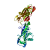
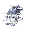
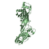
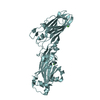
SHEET THE SHEET STRUCTURE OF THIS MOLECULE IS BIFURCATED. IN ORDER TO REPRESENT THIS FEATURE IN ... SHEET THE SHEET STRUCTURE OF THIS MOLECULE IS BIFURCATED. IN ORDER TO REPRESENT THIS FEATURE IN THE SHEET RECORDS BELOW, TWO SHEETS ARE DEFINED.

-
Structure visualization

Structure viewerMolecule:
MolmilJmol/JSmol

Downloads & links

-
Assembly

Deposited unit
A: AGGLUTININ RECEPTOR
hetero molecules


Theoretical massNumber of molelcules
Total (without water)39,8964
Polymers39,7921
Non-polymers1043
Water5,296294
1


  • Idetical with deposited unit
  • defined by author&software
TypeNameSymmetry operationNumber
identity operation1_555x,y,z1
MethodPISA
Unit cell
Length a, b, c (Å)37.198, 61.011, 74.040
Angle α, β, γ (deg.)90.00, 100.09, 90.00
Int Tables number4
Space group name H-MP1211

-
Components

#1: Protein AGGLUTININ RECEPTOR / SSP-5 / SSPB


Mass: 39791.734 Da / Num. of mol.: 1 / Fragment: C-TERMINAL DOMAIN, RESIDUES 1083-1413
Source method: isolated from a genetically manipulated source
Details: ISOPEPTIDE BOND BETWEEN LYS 1259 AND ASN 1393 / Source: (gene. exp.) STREPTOCOCCUS GORDONII (bacteria) / Strain: M5 / Production host: ESCHERICHIA COLI (E. coli) / Strain (production host): BL21(DE3) / References: UniProt: P16952
#2: Chemical ChemComp-CA / CALCIUM ION


Mass: 40.078 Da / Num. of mol.: 2 / Source method: obtained synthetically / Formula: Ca
#3: Chemical ChemComp-MG / MAGNESIUM ION


Mass: 24.305 Da / Num. of mol.: 1 / Source method: obtained synthetically / Formula: Mg
#4: Water ChemComp-HOH / water


Mass: 18.015 Da / Num. of mol.: 294 / Source method: isolated from a natural source / Formula: H2O

-
Experimental details

-
Experiment

ExperimentMethod: X-RAY DIFFRACTION / Number of used crystals: 1

-
Sample preparation

CrystalDensity Matthews: 2.2 Å3/Da / Density % sol: 45 % / Description: NONE
Crystal growpH: 8.5
Details: 0.2 M MGCL2, 0.1 M TRIS-HCL, 30%(W/V) PEG4000, pH 8.5

-
Data collection

DiffractionMean temperature: 100 K
Diffraction sourceSource: SYNCHROTRON / Site: ESRF / Beamline: ID23-1 / Wavelength: 1
DetectorType: ADSC CCD / Detector: CCD / Date: Feb 9, 2009
RadiationMonochromator: SI (111) / Protocol: SINGLE WAVELENGTH / Monochromatic (M) / Laue (L): M / Scattering type: x-ray
Radiation wavelengthWavelength: 1 Å / Relative weight: 1
ReflectionResolution: 1.7→46.78 Å / Num. obs: 33858 / % possible obs: 94.4 % / Observed criterion σ(I): -3 / Redundancy: 2 % / Biso Wilson estimate: 12.38 Å2 / Rmerge(I) obs: 0.06 / Net I/σ(I): 12
Reflection shellResolution: 1.7→1.79 Å / Redundancy: 2 % / Rmerge(I) obs: 0.22 / Mean I/σ(I) obs: 3 / % possible all: 94.6

-
Processing

Software
NameVersionClassification
PHENIX(PHENIX.REFINE)refinement
MOSFLMdata reduction
SCALAdata scaling
MOLREPphasing
RefinementMethod to determine structure: MOLECULAR REPLACEMENT
Starting model: PDB ENTRY 2WOY
Resolution: 1.7→36.623 Å / SU ML: 0.22 / σ(F): 0.01 / Phase error: 22.14 / Stereochemistry target values: ML
Details: A COVALENT BOND IS FORMED BETWEEN THE LYS 1259 NZ AND ASN 1393 CG, SO THE ASN NH2 IS LOST.
RfactorNum. reflection% reflection
Rfree0.2283 1647 5 %
Rwork0.1857 --
obs0.1879 32774 91.11 %
Solvent computationShrinkage radii: 0.9 Å / VDW probe radii: 1.11 Å / Solvent model: FLAT BULK SOLVENT MODEL / Bsol: 31.217 Å2 / ksol: 0.376 e/Å3
Displacement parametersBiso mean: 16.1 Å2
Baniso -1Baniso -2Baniso -3
1-0.7173 Å2-0 Å22.2857 Å2
2--2.7785 Å20 Å2
3----3.3355 Å2
Refinement stepCycle: LAST / Resolution: 1.7→36.623 Å
ProteinNucleic acidLigandSolventTotal
Num. atoms2613 0 3 294 2910
Refine LS restraints
Refine-IDTypeDev idealNumber
X-RAY DIFFRACTIONf_bond_d0.0112664
X-RAY DIFFRACTIONf_angle_d1.3683616
X-RAY DIFFRACTIONf_dihedral_angle_d17.047958
X-RAY DIFFRACTIONf_chiral_restr0.098408
X-RAY DIFFRACTIONf_plane_restr0.006474
LS refinement shell
Resolution (Å)Rfactor RfreeNum. reflection RfreeRfactor RworkNum. reflection RworkRefine-ID% reflection obs (%)
1.7-1.75010.2761160.20372392X-RAY DIFFRACTION85
1.7501-1.80650.25941360.19552433X-RAY DIFFRACTION86
1.8065-1.87110.25831390.18672521X-RAY DIFFRACTION89
1.8711-1.9460.21821170.182554X-RAY DIFFRACTION90
1.946-2.03460.22721350.17662646X-RAY DIFFRACTION93
2.0346-2.14180.24481460.17452620X-RAY DIFFRACTION93
2.1418-2.2760.20241410.18162665X-RAY DIFFRACTION93
2.276-2.45170.26591400.19282696X-RAY DIFFRACTION94
2.4517-2.69840.26861380.2052645X-RAY DIFFRACTION94
2.6984-3.08860.23361560.20232663X-RAY DIFFRACTION93
3.0886-3.89070.2041310.17582606X-RAY DIFFRACTION91
3.8907-36.6310.17631520.15662686X-RAY DIFFRACTION92
Refinement TLS params.

Method: refined / Refine-ID: X-RAY DIFFRACTION

IDL112)L122)L132)L222)L232)L332)S11 (Å °)S12 (Å °)S13 (Å °)S21 (Å °)S22 (Å °)S23 (Å °)S31 (Å °)S32 (Å °)S33 (Å °)T112)T122)T132)T222)T232)T332)Origin x (Å)Origin y (Å)Origin z (Å)
10.4330.081-0.11570.28170.01660.40370.0086-0.00230.0294-0.0293-0.00090.00970.00010.0205-0.01110.0687-0.00190.00310.07390.01970.0568-5.30350.283630.8765
20.3740.0859-0.12890.321-0.130.48380.01010.0123-0.02280.0273-0.00750.0214-0.010.0291-0.00040.04160.0063-0.0110.0328-0.00580.057410.0187-7.8541-7.552
Refinement TLS group
IDRefine-IDRefine TLS-IDSelection details
1X-RAY DIFFRACTION1RESID 1083:1253
2X-RAY DIFFRACTION2RESID 1254:1413

+
About Yorodumi

-
News

-
Feb 9, 2022. New format data for meta-information of EMDB entries

New format data for meta-information of EMDB entries

  • Version 3 of the EMDB header file is now the official format.
  • The previous official version 1.9 will be removed from the archive.

Related info.:EMDB header

External links:wwPDB to switch to version 3 of the EMDB data model

-
Aug 12, 2020. Covid-19 info

Covid-19 info

URL: https://pdbjlvh1.pdbj.org/emnavi/covid19.php

New page: Covid-19 featured information page in EM Navigator.

Related info.:Covid-19 info / Mar 5, 2020. Novel coronavirus structure data

+
Mar 5, 2020. Novel coronavirus structure data

Novel coronavirus structure data

Related info.:Yorodumi Speices / Aug 12, 2020. Covid-19 info

External links:COVID-19 featured content - PDBj / Molecule of the Month (242):Coronavirus Proteases

+
Jan 31, 2019. EMDB accession codes are about to change! (news from PDBe EMDB page)

EMDB accession codes are about to change! (news from PDBe EMDB page)

  • The allocation of 4 digits for EMDB accession codes will soon come to an end. Whilst these codes will remain in use, new EMDB accession codes will include an additional digit and will expand incrementally as the available range of codes is exhausted. The current 4-digit format prefixed with “EMD-” (i.e. EMD-XXXX) will advance to a 5-digit format (i.e. EMD-XXXXX), and so on. It is currently estimated that the 4-digit codes will be depleted around Spring 2019, at which point the 5-digit format will come into force.
  • The EM Navigator/Yorodumi systems omit the EMD- prefix.

Related info.:Q: What is EMD? / ID/Accession-code notation in Yorodumi/EM Navigator

External links:EMDB Accession Codes are Changing Soon! / Contact to PDBj

+
Jul 12, 2017. Major update of PDB

Major update of PDB

  • wwPDB released updated PDB data conforming to the new PDBx/mmCIF dictionary.
  • This is a major update changing the version number from 4 to 5, and with Remediation, in which all the entries are updated.
  • In this update, many items about electron microscopy experimental information are reorganized (e.g. em_software).
  • Now, EM Navigator and Yorodumi are based on the updated data.

External links:wwPDB Remediation / Enriched Model Files Conforming to OneDep Data Standards Now Available in the PDB FTP Archive

-
Yorodumi

Thousand views of thousand structures

  • Yorodumi is a browser for structure data from EMDB, PDB, SASBDB, etc.
  • This page is also the successor to EM Navigator detail page, and also detail information page/front-end page for Omokage search.
  • The word "yorodu" (or yorozu) is an old Japanese word meaning "ten thousand". "mi" (miru) is to see.

Related info.:EMDB / PDB / SASBDB / Comparison of 3 databanks / Yorodumi Search / Aug 31, 2016. New EM Navigator & Yorodumi / Yorodumi Papers / Jmol/JSmol / Function and homology information / Changes in new EM Navigator and Yorodumi

Read more