[English] 日本語
Yorodumi
- PDB-2wd6: Crystal structure of the Variable Domain of the Streptococcus gor... -

+
Open data


ID or keywords:

Loading...

-
Basic information

Entry
Database: PDB / ID: 2wd6
TitleCrystal structure of the Variable Domain of the Streptococcus gordonii Surface Protein SspB
ComponentsAGGLUTININ RECEPTOR
KeywordsCELL ADHESION / SECRETED / V-REGION / CELL WALL / AG I/II PROTEIN / SURFACE ADHESIN / PEPTIDOGLYCAN-ANCHOR
Function / homology
Function and homology information


extracellular region / metal ion binding
Similarity search - Function
Major cell-surface adhesin PAc / Major cell-surface adhesin PAc / Cell surface antigen I/II A repeat / Streptococcal surface antigen repeat / Streptococcus antigen I/II alanine-rich (Ag I/II A) repeat profile. / Glucan-binding protein C/Surface antigen I/II, V-domain / Glucan-binding protein C/Surface antigen I/II, V-domain superfamily / Glucan-binding protein C / Cross-wall-targeting lipoprotein motif / Adhesin isopeptide-forming adherence domain ...Major cell-surface adhesin PAc / Major cell-surface adhesin PAc / Cell surface antigen I/II A repeat / Streptococcal surface antigen repeat / Streptococcus antigen I/II alanine-rich (Ag I/II A) repeat profile. / Glucan-binding protein C/Surface antigen I/II, V-domain / Glucan-binding protein C/Surface antigen I/II, V-domain superfamily / Glucan-binding protein C / Cross-wall-targeting lipoprotein motif / Adhesin isopeptide-forming adherence domain / Cell surface antigen, C-terminal / Antigen I/II, N-terminal / Cell surface antigen C-terminus / Cell surface antigen I/II C2 terminal domain / Adhesin P1 N-terminal domain / LPXTG cell wall anchor motif / Gram-positive cocci surface proteins LPxTG motif profile. / LPXTG cell wall anchor domain / Sandwich / Mainly Beta
Similarity search - Domain/homology
: / Agglutinin receptor
Similarity search - Component
Biological speciesSTREPTOCOCCUS GORDONII (bacteria)
MethodX-RAY DIFFRACTION / SYNCHROTRON / SAD / Resolution: 2.3 Å
AuthorsForsgren, N. / Persson, K.
CitationJournal: Protein Sci. / Year: 2009
Title: Crystal Structure of the Variable Domain of the Streptococcus Gordonii Surface Protein Sspb.
Authors: Forsgren, N. / Lamont, R.J. / Persson, K.
History
DepositionMar 20, 2009Deposition site: PDBE / Processing site: PDBE
Revision 1.0Jul 28, 2009Provider: repository / Type: Initial release
Revision 1.1Jul 13, 2011Group: Advisory / Version format compliance
Revision 1.2Jan 17, 2018Group: Data collection / Category: diffrn_source / Item: _diffrn_source.pdbx_synchrotron_site
Revision 1.3May 8, 2024Group: Data collection / Database references ...Data collection / Database references / Derived calculations / Other
Category: chem_comp_atom / chem_comp_bond ...chem_comp_atom / chem_comp_bond / database_2 / pdbx_database_status / pdbx_struct_conn_angle / struct_conn / struct_site
Item: _database_2.pdbx_DOI / _database_2.pdbx_database_accession ..._database_2.pdbx_DOI / _database_2.pdbx_database_accession / _pdbx_database_status.status_code_sf / _pdbx_struct_conn_angle.ptnr1_auth_asym_id / _pdbx_struct_conn_angle.ptnr1_auth_comp_id / _pdbx_struct_conn_angle.ptnr1_auth_seq_id / _pdbx_struct_conn_angle.ptnr1_label_asym_id / _pdbx_struct_conn_angle.ptnr1_label_atom_id / _pdbx_struct_conn_angle.ptnr1_label_comp_id / _pdbx_struct_conn_angle.ptnr1_label_seq_id / _pdbx_struct_conn_angle.ptnr3_auth_asym_id / _pdbx_struct_conn_angle.ptnr3_auth_comp_id / _pdbx_struct_conn_angle.ptnr3_auth_seq_id / _pdbx_struct_conn_angle.ptnr3_label_asym_id / _pdbx_struct_conn_angle.ptnr3_label_atom_id / _pdbx_struct_conn_angle.ptnr3_label_comp_id / _pdbx_struct_conn_angle.ptnr3_label_seq_id / _pdbx_struct_conn_angle.value / _struct_conn.pdbx_dist_value / _struct_conn.ptnr1_auth_asym_id / _struct_conn.ptnr1_auth_comp_id / _struct_conn.ptnr1_auth_seq_id / _struct_conn.ptnr1_label_asym_id / _struct_conn.ptnr1_label_atom_id / _struct_conn.ptnr1_label_comp_id / _struct_conn.ptnr1_label_seq_id / _struct_conn.ptnr2_auth_asym_id / _struct_conn.ptnr2_auth_comp_id / _struct_conn.ptnr2_auth_seq_id / _struct_conn.ptnr2_label_asym_id / _struct_conn.ptnr2_label_atom_id / _struct_conn.ptnr2_label_comp_id / _struct_conn.ptnr2_label_seq_id / _struct_site.pdbx_auth_asym_id / _struct_site.pdbx_auth_comp_id / _struct_site.pdbx_auth_seq_id
Remark 700 
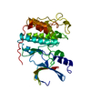
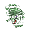
SHEET THE SHEET STRUCTURE OF THIS MOLECULE IS BIFURCATED. IN ORDER TO REPRESENT THIS FEATURE IN ... SHEET THE SHEET STRUCTURE OF THIS MOLECULE IS BIFURCATED. IN ORDER TO REPRESENT THIS FEATURE IN THE SHEET RECORDS BELOW, TWO SHEETS ARE DEFINED.

-
Structure visualization

Structure viewerMolecule:
MolmilJmol/JSmol

Downloads & links

-
Assembly

Deposited unit
A: AGGLUTININ RECEPTOR
B: AGGLUTININ RECEPTOR
hetero molecules


Theoretical massNumber of molelcules
Total (without water)77,5419
Polymers77,0532
Non-polymers4887
Water5,062281
1
A: AGGLUTININ RECEPTOR
hetero molecules


Theoretical massNumber of molelcules
Total (without water)38,7514
Polymers38,5271
Non-polymers2243
Water181
TypeNameSymmetry operationNumber
identity operation1_555x,y,z1
2
B: AGGLUTININ RECEPTOR
hetero molecules


Theoretical massNumber of molelcules
Total (without water)38,7905
Polymers38,5271
Non-polymers2634
Water181
TypeNameSymmetry operationNumber
identity operation1_555x,y,z1
3


  • Idetical with deposited unit
  • defined by software
TypeNameSymmetry operationNumber
identity operation1_555x,y,z1
Buried area3610 Å2
ΔGint-8.1 kcal/mol
Surface area28380 Å2
MethodPQS
Unit cell
Length a, b, c (Å)110.170, 110.170, 121.490
Angle α, β, γ (deg.)90.00, 90.00, 120.00
Int Tables number170
Space group name H-MP65

-
Components

#1: Protein AGGLUTININ RECEPTOR / SSP-5 / CELL SURFACE ANTIGEN


Mass: 38526.527 Da / Num. of mol.: 2 / Fragment: VARIABLE DOMAIN, RESIDUES 449-797
Source method: isolated from a genetically manipulated source
Source: (gene. exp.) STREPTOCOCCUS GORDONII (bacteria) / Strain: M5 / Production host: ESCHERICHIA COLI (E. coli) / Strain (production host): BL21(DE3) / References: UniProt: P16952
#2: Chemical ChemComp-CA / CALCIUM ION


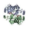
Mass: 40.078 Da / Num. of mol.: 2 / Source method: obtained synthetically / Formula: Ca
#3: Chemical
ChemComp-GOL / GLYCEROL / GLYCERIN / PROPANE-1,2,3-TRIOL


Mass: 92.094 Da / Num. of mol.: 4 / Source method: obtained synthetically / Formula: C3H8O3
#4: Chemical ChemComp-K / POTASSIUM ION


Mass: 39.098 Da / Num. of mol.: 1 / Source method: obtained synthetically / Formula: K
#5: Water ChemComp-HOH / water


Mass: 18.015 Da / Num. of mol.: 281 / Source method: isolated from a natural source / Formula: H2O
Nonpolymer detailsPOTASSIUM ION (K): POSITIONED BETWEEN CHAIN A AND CHAIN B GLYCEROL (GOL): TWO GLYCEROL MOLECULES ...POTASSIUM ION (K): POSITIONED BETWEEN CHAIN A AND CHAIN B GLYCEROL (GOL): TWO GLYCEROL MOLECULES ARE MODELLED IN THE CAVITY CLOSE TO THE CALCIUM ION
Sequence detailsTHIS IS THE SEQUENCE FOR THE WHOLE SSPB PROTEIN SINCE THERE IS NO SPECIFIC ENTRY FOR ONLY THE V-DOMAIN ITSELF

-
Experimental details

-
Experiment

ExperimentMethod: X-RAY DIFFRACTION / Number of used crystals: 1

-
Sample preparation

CrystalDensity Matthews: 2.8 Å3/Da / Density % sol: 55 % / Description: NONE
Crystal growDetails: 0.2 M KBR 25% PEG3350

-
Data collection

DiffractionMean temperature: 100 K
Diffraction sourceSource: SYNCHROTRON / Site: MAX II / Beamline: I911-3 / Wavelength: 0.91698
DetectorType: MARMOSAIC 225 mm CCD / Detector: CCD / Date: Oct 27, 2007
RadiationMonochromator: SI(111) / Protocol: SINGLE WAVELENGTH / Monochromatic (M) / Laue (L): M / Scattering type: x-ray
Radiation wavelengthWavelength: 0.91698 Å / Relative weight: 1
ReflectionResolution: 2.3→60.75 Å / Num. obs: 37172 / % possible obs: 100 % / Observed criterion σ(I): 2 / Redundancy: 10.99 % / Rmerge(I) obs: 0.1 / Net I/σ(I): 24.4
Reflection shellResolution: 2.3→2.42 Å / Redundancy: 10.42 % / Rmerge(I) obs: 0.52 / Mean I/σ(I) obs: 4.4 / % possible all: 99.8

-
Processing

Software
NameVersionClassification
REFMAC5.2.0019refinement
MOSFLMdata reduction
SCALAdata scaling
PHENIXphasing
RefinementMethod to determine structure: SAD
Starting model: NONE

Resolution: 2.3→95.35 Å / Cor.coef. Fo:Fc: 0.947 / Cor.coef. Fo:Fc free: 0.929 / SU B: 9.743 / SU ML: 0.128 / TLS residual ADP flag: LIKELY RESIDUAL / Cross valid method: THROUGHOUT / ESU R: 0.233 / ESU R Free: 0.183 / Stereochemistry target values: MAXIMUM LIKELIHOOD / Details: HYDROGENS HAVE BEEN ADDED IN THE RIDING POSITIONS.
RfactorNum. reflection% reflectionSelection details
Rfree0.209 1853 5 %RANDOM
Rwork0.176 ---
obs0.177 35258 99.9 %-
Solvent computationIon probe radii: 0.8 Å / Shrinkage radii: 0.8 Å / VDW probe radii: 1.2 Å / Solvent model: MASK
Displacement parametersBiso mean: 24.84 Å2
Baniso -1Baniso -2Baniso -3
1--0.21 Å2-0.1 Å20 Å2
2---0.21 Å20 Å2
3---0.31 Å2
Refinement stepCycle: LAST / Resolution: 2.3→95.35 Å
ProteinNucleic acidLigandSolventTotal
Num. atoms4638 0 27 281 4946
Refine LS restraints
Refine-IDTypeDev idealDev ideal targetNumber
X-RAY DIFFRACTIONr_bond_refined_d0.020.0224761
X-RAY DIFFRACTIONr_bond_other_d
X-RAY DIFFRACTIONr_angle_refined_deg1.6341.9346444
X-RAY DIFFRACTIONr_angle_other_deg
X-RAY DIFFRACTIONr_dihedral_angle_1_deg7.2375593
X-RAY DIFFRACTIONr_dihedral_angle_2_deg38.63325.294221
X-RAY DIFFRACTIONr_dihedral_angle_3_deg16.36815789
X-RAY DIFFRACTIONr_dihedral_angle_4_deg17.8131516
X-RAY DIFFRACTIONr_chiral_restr0.1070.2690
X-RAY DIFFRACTIONr_gen_planes_refined0.0060.023635
X-RAY DIFFRACTIONr_gen_planes_other
X-RAY DIFFRACTIONr_nbd_refined0.210.22046
X-RAY DIFFRACTIONr_nbd_other
X-RAY DIFFRACTIONr_nbtor_refined0.3050.23179
X-RAY DIFFRACTIONr_nbtor_other
X-RAY DIFFRACTIONr_xyhbond_nbd_refined0.1560.2366
X-RAY DIFFRACTIONr_xyhbond_nbd_other
X-RAY DIFFRACTIONr_metal_ion_refined
X-RAY DIFFRACTIONr_metal_ion_other
X-RAY DIFFRACTIONr_symmetry_vdw_refined0.1660.222
X-RAY DIFFRACTIONr_symmetry_vdw_other
X-RAY DIFFRACTIONr_symmetry_hbond_refined0.110.23
X-RAY DIFFRACTIONr_symmetry_hbond_other
X-RAY DIFFRACTIONr_symmetry_metal_ion_refined
X-RAY DIFFRACTIONr_symmetry_metal_ion_other
X-RAY DIFFRACTIONr_mcbond_it0.6681.52954
X-RAY DIFFRACTIONr_mcbond_other
X-RAY DIFFRACTIONr_mcangle_it1.23724758
X-RAY DIFFRACTIONr_mcangle_other
X-RAY DIFFRACTIONr_scbond_it2.36331839
X-RAY DIFFRACTIONr_scbond_other
X-RAY DIFFRACTIONr_scangle_it3.6884.51686
X-RAY DIFFRACTIONr_scangle_other
X-RAY DIFFRACTIONr_long_range_B_refined
X-RAY DIFFRACTIONr_long_range_B_other
X-RAY DIFFRACTIONr_rigid_bond_restr
X-RAY DIFFRACTIONr_sphericity_free
X-RAY DIFFRACTIONr_sphericity_bonded
LS refinement shellResolution: 2.3→2.36 Å / Total num. of bins used: 20
RfactorNum. reflection% reflection
Rfree0.257 123 -
Rwork0.204 2606 -
obs--99.78 %
Refinement TLS params.

Method: refined / Refine-ID: X-RAY DIFFRACTION

IDL112)L122)L132)L222)L232)L332)S11 (Å °)S12 (Å °)S13 (Å °)S21 (Å °)S22 (Å °)S23 (Å °)S31 (Å °)S32 (Å °)S33 (Å °)T112)T122)T132)T222)T232)T332)Origin x (Å)Origin y (Å)Origin z (Å)
11.5550.10820.11711.3868-0.12632.2578-0.0555-0.04920.1005-0.02420.0207-0.2081-0.09410.0610.0348-0.15210.0226-0.0121-0.1276-0.0072-0.009941.728638.447875.5783
23.6299-0.60880.85612.19740.31272.3527-0.1211-0.6430.15280.07580.15610.16490.0019-0.2262-0.035-0.14090.0765-0.02550.0636-0.0401-0.037116.249941.358577.9319
Refinement TLS group
IDRefine-IDRefine TLS-IDAuth asym-IDAuth seq-ID
1X-RAY DIFFRACTION1A462 - 762
2X-RAY DIFFRACTION2B467 - 760

+
About Yorodumi

-
News

-
Feb 9, 2022. New format data for meta-information of EMDB entries

New format data for meta-information of EMDB entries

  • Version 3 of the EMDB header file is now the official format.
  • The previous official version 1.9 will be removed from the archive.

Related info.:EMDB header

External links:wwPDB to switch to version 3 of the EMDB data model

-
Aug 12, 2020. Covid-19 info

Covid-19 info

URL: https://pdbjlvh1.pdbj.org/emnavi/covid19.php

New page: Covid-19 featured information page in EM Navigator.

Related info.:Covid-19 info / Mar 5, 2020. Novel coronavirus structure data

+
Mar 5, 2020. Novel coronavirus structure data

Novel coronavirus structure data

Related info.:Yorodumi Speices / Aug 12, 2020. Covid-19 info

External links:COVID-19 featured content - PDBj / Molecule of the Month (242):Coronavirus Proteases

+
Jan 31, 2019. EMDB accession codes are about to change! (news from PDBe EMDB page)

EMDB accession codes are about to change! (news from PDBe EMDB page)

  • The allocation of 4 digits for EMDB accession codes will soon come to an end. Whilst these codes will remain in use, new EMDB accession codes will include an additional digit and will expand incrementally as the available range of codes is exhausted. The current 4-digit format prefixed with “EMD-” (i.e. EMD-XXXX) will advance to a 5-digit format (i.e. EMD-XXXXX), and so on. It is currently estimated that the 4-digit codes will be depleted around Spring 2019, at which point the 5-digit format will come into force.
  • The EM Navigator/Yorodumi systems omit the EMD- prefix.

Related info.:Q: What is EMD? / ID/Accession-code notation in Yorodumi/EM Navigator

External links:EMDB Accession Codes are Changing Soon! / Contact to PDBj

+
Jul 12, 2017. Major update of PDB

Major update of PDB

  • wwPDB released updated PDB data conforming to the new PDBx/mmCIF dictionary.
  • This is a major update changing the version number from 4 to 5, and with Remediation, in which all the entries are updated.
  • In this update, many items about electron microscopy experimental information are reorganized (e.g. em_software).
  • Now, EM Navigator and Yorodumi are based on the updated data.

External links:wwPDB Remediation / Enriched Model Files Conforming to OneDep Data Standards Now Available in the PDB FTP Archive

-
Yorodumi

Thousand views of thousand structures

  • Yorodumi is a browser for structure data from EMDB, PDB, SASBDB, etc.
  • This page is also the successor to EM Navigator detail page, and also detail information page/front-end page for Omokage search.
  • The word "yorodu" (or yorozu) is an old Japanese word meaning "ten thousand". "mi" (miru) is to see.

Related info.:EMDB / PDB / SASBDB / Comparison of 3 databanks / Yorodumi Search / Aug 31, 2016. New EM Navigator & Yorodumi / Yorodumi Papers / Jmol/JSmol / Function and homology information / Changes in new EM Navigator and Yorodumi

Read more