[English] 日本語
Yorodumi
- PDB-2wyi: Structure of the Streptococcus pyogenes family GH38 alpha-mannosi... -

+
Open data


ID or keywords:

Loading...

-
Basic information

Entry
Database: PDB / ID: 2wyi
Title
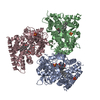
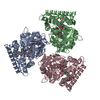
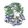
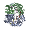
Structure of the Streptococcus pyogenes family GH38 alpha-mannosidase complexed with swainsonine
ComponentsALPHA-MANNOSIDASE
KeywordsHYDROLASE / GLYCOSIDASE / GLYCOSIDE HYDROLASE
Function / homology
Function and homology information


alpha-mannosidase activity / mannose metabolic process / oligosaccharide catabolic process / carbohydrate binding / metal ion binding
Similarity search - Function
Methyltransferase, Methionine Synthase (B12-binding Domains); Chain A, domain 1 - #90 / Rossmann fold - #11160 / Immunoglobulin-like - #2210 / Immunoglobulin-like - #2220 / Glycosyl hydrolases 38, beta-1 domain / Glycosyl hydrolases family 38 C-terminal domain 1 / Glycosyl hydrolases family 38, C-terminal beta sandwich domain / Glycosyl hydrolases family 38 C-terminal beta sandwich domain / Methyltransferase, Methionine Synthase (B12-binding Domains); Chain A, domain 1 / Glycoside hydrolase family 38, central domain ...Methyltransferase, Methionine Synthase (B12-binding Domains); Chain A, domain 1 - #90 / Rossmann fold - #11160 / Immunoglobulin-like - #2210 / Immunoglobulin-like - #2220 / Glycosyl hydrolases 38, beta-1 domain / Glycosyl hydrolases family 38 C-terminal domain 1 / Glycosyl hydrolases family 38, C-terminal beta sandwich domain / Glycosyl hydrolases family 38 C-terminal beta sandwich domain / Methyltransferase, Methionine Synthase (B12-binding Domains); Chain A, domain 1 / Glycoside hydrolase family 38, central domain / Golgi alpha-mannosidase II; domain 4 / Glycoside hydrolase family 38, N-terminal domain / Glycosyl hydrolase family 38, C-terminal / Glycoside hydrolase family 38, central domain / Glycoside hydrolase family 38, central domain superfamily / Glycosyl hydrolases family 38 N-terminal domain / Glycosyl hydrolases family 38 C-terminal domain / Alpha mannosidase middle domain / Alpha mannosidase, middle domain / Glycoside hydrolase 38, N-terminal domain superfamily / Glycoside hydrolase families 57/38, central domain superfamily / Glycoside hydrolase/deacetylase, beta/alpha-barrel / Substrate Binding Domain Of Dnak; Chain:A; Domain 2 / Beta-galactosidase; Chain A, domain 5 / Galactose mutarotase-like domain superfamily / Distorted Sandwich / Up-down Bundle / Immunoglobulin-like / Sandwich / Rossmann fold / Orthogonal Bundle / 3-Layer(aba) Sandwich / Mainly Beta / Mainly Alpha / Alpha Beta
Similarity search - Domain/homology
1S-8AB-OCTAHYDRO-INDOLIZIDINE-1A,2A,8B-TRIOL / Glycoside hydrolase family 38 central domain-containing protein
Similarity search - Component
Biological speciesSTREPTOCOCCUS PYOGENES (bacteria)
MethodX-RAY DIFFRACTION / SYNCHROTRON / MAD / Resolution: 2.6 Å
AuthorsSuits, M.D.L. / Zhu, Y. / Taylor, E.J. / Zechel, D.L. / Gilbert, H.J. / Davies, G.J.
CitationJournal: Plos One / Year: 2010
Title: Structure and Kinetic Investigation of Streptococcus Pyogenes Family Gh38 Alpha-Mannosidase
Authors: Suits, M.D.L. / Zhu, Y. / Taylor, E.J. / Zechel, D.L. / Gilbert, H.J. / Davies, G.J.
History
DepositionNov 16, 2009Deposition site: PDBE / Processing site: PDBE
Revision 1.0Feb 16, 2010Provider: repository / Type: Initial release
Revision 1.1Jul 13, 2011Group: Advisory / Version format compliance
Revision 1.2Dec 20, 2023Group: Data collection / Database references ...Data collection / Database references / Derived calculations / Other / Refinement description
Category: chem_comp_atom / chem_comp_bond ...chem_comp_atom / chem_comp_bond / database_2 / pdbx_database_status / pdbx_initial_refinement_model / pdbx_struct_conn_angle / struct_conn / struct_site
Item: _database_2.pdbx_DOI / _database_2.pdbx_database_accession ..._database_2.pdbx_DOI / _database_2.pdbx_database_accession / _pdbx_database_status.status_code_sf / _pdbx_struct_conn_angle.ptnr1_auth_comp_id / _pdbx_struct_conn_angle.ptnr1_auth_seq_id / _pdbx_struct_conn_angle.ptnr1_label_asym_id / _pdbx_struct_conn_angle.ptnr1_label_atom_id / _pdbx_struct_conn_angle.ptnr1_label_comp_id / _pdbx_struct_conn_angle.ptnr1_label_seq_id / _pdbx_struct_conn_angle.ptnr3_auth_comp_id / _pdbx_struct_conn_angle.ptnr3_auth_seq_id / _pdbx_struct_conn_angle.ptnr3_label_asym_id / _pdbx_struct_conn_angle.ptnr3_label_atom_id / _pdbx_struct_conn_angle.ptnr3_label_comp_id / _pdbx_struct_conn_angle.ptnr3_label_seq_id / _pdbx_struct_conn_angle.value / _struct_conn.pdbx_dist_value / _struct_conn.ptnr1_auth_comp_id / _struct_conn.ptnr1_auth_seq_id / _struct_conn.ptnr1_label_asym_id / _struct_conn.ptnr1_label_atom_id / _struct_conn.ptnr1_label_comp_id / _struct_conn.ptnr1_label_seq_id / _struct_conn.ptnr2_auth_comp_id / _struct_conn.ptnr2_auth_seq_id / _struct_conn.ptnr2_label_asym_id / _struct_conn.ptnr2_label_atom_id / _struct_conn.ptnr2_label_comp_id / _struct_conn.ptnr2_label_seq_id / _struct_site.pdbx_auth_asym_id / _struct_site.pdbx_auth_comp_id / _struct_site.pdbx_auth_seq_id

-
Structure visualization

Structure viewerMolecule:
MolmilJmol/JSmol

Downloads & links

-
Assembly

Deposited unit
A: ALPHA-MANNOSIDASE
B: ALPHA-MANNOSIDASE
hetero molecules


Theoretical massNumber of molelcules
Total (without water)212,28217
Polymers210,4832
Non-polymers1,79915
Water6,215345
1


  • Idetical with deposited unit
  • defined by author&software
TypeNameSymmetry operationNumber
identity operation1_555x,y,z1
Buried area9790 Å2
ΔGint-46.1 kcal/mol
Surface area62150 Å2
MethodPISA
Unit cell
Length a, b, c (Å)178.680, 178.680, 198.237
Angle α, β, γ (deg.)90.00, 90.00, 90.00
Int Tables number96
Space group name H-MP43212
Noncrystallographic symmetry (NCS)NCS domain:
IDEns-IDDetails
11A
21B
12A
22B

NCS domain segments:
Dom-IDComponent-IDEns-IDRefine codeAuth asym-IDAuth seq-ID
1115A1 - 901
2115B3 - 901
1124A1000 - 1001
2124B1000 - 1001

NCS ensembles :
ID
1
2

-
Components

#1: Protein ALPHA-MANNOSIDASE / ALPHA-MANNOSIDASE II


Mass: 105241.656 Da / Num. of mol.: 2
Source method: isolated from a genetically manipulated source
Source: (gene. exp.) STREPTOCOCCUS PYOGENES (bacteria) / Strain: M1 GAS / Production host: ESCHERICHIA COLI (E. coli) / Strain (production host): BL21(DE3) / References: UniProt: Q99YP5, alpha-mannosidase
#2: Chemical
ChemComp-PG0 / 2-(2-METHOXYETHOXY)ETHANOL / PEG 6000


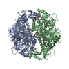
Mass: 120.147 Da / Num. of mol.: 11 / Source method: obtained synthetically / Formula: C5H12O3 / Comment: inhibitor, precipitant*YM
#3: Chemical ChemComp-ZN / ZINC ION


Mass: 65.409 Da / Num. of mol.: 2 / Source method: obtained synthetically / Formula: Zn
#4: Chemical ChemComp-SWA / 1S-8AB-OCTAHYDRO-INDOLIZIDINE-1A,2A,8B-TRIOL / SWAINSONINE


Mass: 173.210 Da / Num. of mol.: 2 / Source method: obtained synthetically / Formula: C8H15NO3 / Comment: chemotherapy, inhibitor, alkaloid*YM
#5: Water ChemComp-HOH / water


Mass: 18.015 Da / Num. of mol.: 345 / Source method: isolated from a natural source / Formula: H2O
Sequence detailsTHE FIRST 22 RESIDUES ARE A C3 PROTEASE CLEAVABLE AFFINITY TAG (MGSSHHHHHHSSGLEVLFQGPA)

-
Experimental details

-
Experiment

ExperimentMethod: X-RAY DIFFRACTION / Number of used crystals: 1

-
Sample preparation

CrystalDensity Matthews: 3.84 Å3/Da / Density % sol: 69 % / Description: NONE
Crystal growpH: 7
Details: 15 MG/ML 3C CLEAVED SPGH38 MIXED WITH 3% V/V GLYCEROL, 54% V/V TACSIMATE (PH 7.0) AND 2% V/V POLYETHYLENE GLYCOL 6000

-
Data collection

DiffractionMean temperature: 100 K
Diffraction sourceSource: SYNCHROTRON / Site: ESRF / Beamline: ID14-1 / Wavelength: 0.9334
DetectorType: ADSC CCD / Detector: CCD / Date: Nov 21, 2008
RadiationProtocol: MAD / Monochromatic (M) / Laue (L): M / Scattering type: x-ray
Radiation wavelengthWavelength: 0.9334 Å / Relative weight: 1
ReflectionResolution: 2.6→50 Å / Num. obs: 93181 / % possible obs: 99.8 % / Observed criterion σ(I): 2 / Redundancy: 6.8 % / Rmerge(I) obs: 0.05 / Net I/σ(I): 27.7
Reflection shellResolution: 2.6→2.69 Å / Redundancy: 7 % / Rmerge(I) obs: 0.39 / Mean I/σ(I) obs: 4.1 / % possible all: 100

-
Processing

Software
NameVersionClassification
PHASERmodel building
SCALEPACKdata scaling
SHARPphasing
SHELXphasing
RESOLVEphasing
PHASERphasing
REFMAC5.5.0109refinement
RefinementMethod to determine structure: MAD
Starting model: PDB ENTRY 2WYH
Resolution: 2.6→132.45 Å / Cor.coef. Fo:Fc: 0.949 / Cor.coef. Fo:Fc free: 0.925 / SU B: 18.421 / SU ML: 0.175 / TLS residual ADP flag: LIKELY RESIDUAL / Cross valid method: THROUGHOUT / ESU R: 0.327 / ESU R Free: 0.235 / Stereochemistry target values: MAXIMUM LIKELIHOOD
Details: HYDROGENS HAVE BEEN ADDED IN THE RIDING POSITIONS. U VALUES RESIDUAL ONLY DISORDERED REGION OF MOLECULES WERE MODELED STEREOCHEMICALLY AGAINST THE APO STRUCTURE (RESIDUES 156- 163). ATOM ...Details: HYDROGENS HAVE BEEN ADDED IN THE RIDING POSITIONS. U VALUES RESIDUAL ONLY DISORDERED REGION OF MOLECULES WERE MODELED STEREOCHEMICALLY AGAINST THE APO STRUCTURE (RESIDUES 156- 163). ATOM RECORD CONTAINS RESIDUAL B FACTORS ONLY
RfactorNum. reflection% reflectionSelection details
Rfree0.223 4888 5 %RANDOM
Rwork0.187 ---
obs0.189 93181 99.7 %-
Solvent computationIon probe radii: 0.8 Å / Shrinkage radii: 0.8 Å / VDW probe radii: 1.4 Å / Solvent model: MASK
Displacement parametersBiso mean: 28.09 Å2
Baniso -1Baniso -2Baniso -3
1-0.89 Å20 Å20 Å2
2--0.89 Å20 Å2
3----1.77 Å2
Refinement stepCycle: LAST / Resolution: 2.6→132.45 Å
ProteinNucleic acidLigandSolventTotal
Num. atoms14274 0 114 345 14733
Refine LS restraints
Refine-IDTypeDev idealDev ideal targetNumber
X-RAY DIFFRACTIONr_bond_refined_d0.0090.02214778
X-RAY DIFFRACTIONr_bond_other_d
X-RAY DIFFRACTIONr_angle_refined_deg1.1111.94920061
X-RAY DIFFRACTIONr_angle_other_deg
X-RAY DIFFRACTIONr_dihedral_angle_1_deg5.72451808
X-RAY DIFFRACTIONr_dihedral_angle_2_deg33.95824.501742
X-RAY DIFFRACTIONr_dihedral_angle_3_deg14.68152434
X-RAY DIFFRACTIONr_dihedral_angle_4_deg19.0231588
X-RAY DIFFRACTIONr_chiral_restr0.0750.22201
X-RAY DIFFRACTIONr_gen_planes_refined0.0040.02111361
X-RAY DIFFRACTIONr_gen_planes_other
X-RAY DIFFRACTIONr_nbd_refined
X-RAY DIFFRACTIONr_nbd_other
X-RAY DIFFRACTIONr_nbtor_refined
X-RAY DIFFRACTIONr_nbtor_other
X-RAY DIFFRACTIONr_xyhbond_nbd_refined
X-RAY DIFFRACTIONr_xyhbond_nbd_other
X-RAY DIFFRACTIONr_metal_ion_refined
X-RAY DIFFRACTIONr_metal_ion_other
X-RAY DIFFRACTIONr_symmetry_vdw_refined
X-RAY DIFFRACTIONr_symmetry_vdw_other
X-RAY DIFFRACTIONr_symmetry_hbond_refined
X-RAY DIFFRACTIONr_symmetry_hbond_other
X-RAY DIFFRACTIONr_symmetry_metal_ion_refined
X-RAY DIFFRACTIONr_symmetry_metal_ion_other
X-RAY DIFFRACTIONr_mcbond_it0.3961.58979
X-RAY DIFFRACTIONr_mcbond_other
X-RAY DIFFRACTIONr_mcangle_it0.782214493
X-RAY DIFFRACTIONr_mcangle_other
X-RAY DIFFRACTIONr_scbond_it1.19335799
X-RAY DIFFRACTIONr_scbond_other
X-RAY DIFFRACTIONr_scangle_it2.0414.55558
X-RAY DIFFRACTIONr_scangle_other
X-RAY DIFFRACTIONr_long_range_B_refined
X-RAY DIFFRACTIONr_long_range_B_other
X-RAY DIFFRACTIONr_rigid_bond_restr
X-RAY DIFFRACTIONr_sphericity_free
X-RAY DIFFRACTIONr_sphericity_bonded
Refine LS restraints NCS

Refine-ID: X-RAY DIFFRACTION

Ens-IDDom-IDAuth asym-IDNumberTypeRms dev position (Å)Weight position
11A3572medium positional0.120.5
12B3572medium positional0.120.5
21A13medium positional0.160.5
22B13medium positional0.160.5
11A3490loose positional0.25
12B3490loose positional0.25
11A3572medium thermal0.352
12B3572medium thermal0.352
21A13medium thermal0.32
22B13medium thermal0.32
11A3490loose thermal0.4810
12B3490loose thermal0.4810
LS refinement shellResolution: 2.6→2.67 Å / Total num. of bins used: 20
RfactorNum. reflection% reflection
Rfree0.296 358 -
Rwork0.266 6786 -
obs--99.33 %
Refinement TLS params.

Method: refined / Refine-ID: X-RAY DIFFRACTION

IDL112)L122)L132)L222)L232)L332)S11 (Å °)S12 (Å °)S13 (Å °)S21 (Å °)S22 (Å °)S23 (Å °)S31 (Å °)S32 (Å °)S33 (Å °)T112)T122)T132)T222)T232)T332)Origin x (Å)Origin y (Å)Origin z (Å)
10.7310.2130.13680.4806-0.01380.61550.10010.0618-0.00830.0105-0.1346-0.09760.09490.17890.03450.04310.0489-0.00930.15830.05310.047729.5857-61.333313.3467
20.67370.13550.30710.53540.1770.9816-0.04550.09260.3779-0.0551-0.10150.082-0.29330.03880.1470.158-0.0061-0.04350.13930.10980.258618.5605-23.17576.7078
Refinement TLS group
IDRefine-IDRefine TLS-IDAuth asym-IDAuth seq-ID
1X-RAY DIFFRACTION1A1 - 901
2X-RAY DIFFRACTION2B3 - 901

+
About Yorodumi

-
News

-
Feb 9, 2022. New format data for meta-information of EMDB entries

New format data for meta-information of EMDB entries

  • Version 3 of the EMDB header file is now the official format.
  • The previous official version 1.9 will be removed from the archive.

Related info.:EMDB header

External links:wwPDB to switch to version 3 of the EMDB data model

-
Aug 12, 2020. Covid-19 info

Covid-19 info

URL: https://pdbjlvh1.pdbj.org/emnavi/covid19.php

New page: Covid-19 featured information page in EM Navigator.

Related info.:Covid-19 info / Mar 5, 2020. Novel coronavirus structure data

+
Mar 5, 2020. Novel coronavirus structure data

Novel coronavirus structure data

Related info.:Yorodumi Speices / Aug 12, 2020. Covid-19 info

External links:COVID-19 featured content - PDBj / Molecule of the Month (242):Coronavirus Proteases

+
Jan 31, 2019. EMDB accession codes are about to change! (news from PDBe EMDB page)

EMDB accession codes are about to change! (news from PDBe EMDB page)

  • The allocation of 4 digits for EMDB accession codes will soon come to an end. Whilst these codes will remain in use, new EMDB accession codes will include an additional digit and will expand incrementally as the available range of codes is exhausted. The current 4-digit format prefixed with “EMD-” (i.e. EMD-XXXX) will advance to a 5-digit format (i.e. EMD-XXXXX), and so on. It is currently estimated that the 4-digit codes will be depleted around Spring 2019, at which point the 5-digit format will come into force.
  • The EM Navigator/Yorodumi systems omit the EMD- prefix.

Related info.:Q: What is EMD? / ID/Accession-code notation in Yorodumi/EM Navigator

External links:EMDB Accession Codes are Changing Soon! / Contact to PDBj

+
Jul 12, 2017. Major update of PDB

Major update of PDB

  • wwPDB released updated PDB data conforming to the new PDBx/mmCIF dictionary.
  • This is a major update changing the version number from 4 to 5, and with Remediation, in which all the entries are updated.
  • In this update, many items about electron microscopy experimental information are reorganized (e.g. em_software).
  • Now, EM Navigator and Yorodumi are based on the updated data.

External links:wwPDB Remediation / Enriched Model Files Conforming to OneDep Data Standards Now Available in the PDB FTP Archive

-
Yorodumi

Thousand views of thousand structures

  • Yorodumi is a browser for structure data from EMDB, PDB, SASBDB, etc.
  • This page is also the successor to EM Navigator detail page, and also detail information page/front-end page for Omokage search.
  • The word "yorodu" (or yorozu) is an old Japanese word meaning "ten thousand". "mi" (miru) is to see.

Related info.:EMDB / PDB / SASBDB / Comparison of 3 databanks / Yorodumi Search / Aug 31, 2016. New EM Navigator & Yorodumi / Yorodumi Papers / Jmol/JSmol / Function and homology information / Changes in new EM Navigator and Yorodumi

Read more