- PDB-2wyh: Structure of the Streptococcus pyogenes family GH38 alpha-mannosidase -
+
Open data
ID or keywords:
Loading...
-
Basic information
Entry
Database: PDB / ID: 2wyh
Title
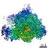
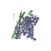
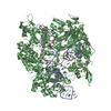
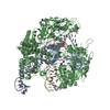
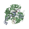
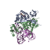
Structure of the Streptococcus pyogenes family GH38 alpha-mannosidase
Components
ALPHA-MANNOSIDASE
Keywords
HYDROLASE / GLYCOSIDASE / GLYCOSIDE HYDROLASE
Function / homology
Function and homology information
alpha-mannosidase activity / mannose metabolic process / oligosaccharide catabolic process / carbohydrate binding / metal ion binding Similarity search - Function
Resolution: 1.9→50 Å / Num. obs: 152396 / % possible obs: 99.4 % / Observed criterion σ(I): 2 / Redundancy: 3.3 % / Rmerge(I) obs: 0.05 / Net I/σ(I): 14.4
Reflection shell
Resolution: 1.9→1.97 Å / Redundancy: 3 % / Rmerge(I) obs: 0.46 / Mean I/σ(I) obs: 1.95 / % possible all: 99.1
-
Processing
Software
Name
Version
Classification
REFMAC
5.5.0109
refinement
HKL-2000
datareduction
SCALEPACK
datascaling
SHARP
phasing
SHELX
phasing
RESOLVE
phasing
Refinement
Method to determine structure: MAD Starting model: NONE Resolution: 1.9→127 Å / Cor.coef. Fo:Fc: 0.953 / Cor.coef. Fo:Fc free: 0.94 / SU B: 6.185 / SU ML: 0.082 / TLS residual ADP flag: LIKELY RESIDUAL / Cross valid method: THROUGHOUT / ESU R: 0.142 / ESU R Free: 0.126 / Stereochemistry target values: MAXIMUM LIKELIHOOD Details: HYDROGENS HAVE BEEN ADDED IN THE RIDING POSITIONS. U VALUES RESIDUAL ONLY. DISORDERED REGION OF MOLECULE B (RESIDUES 156-164).
Rfactor
Num. reflection
% reflection
Selection details
Rfree
0.205
8041
5 %
RANDOM
Rwork
0.177
-
-
-
obs
0.179
152396
99.1 %
-
Solvent computation
Ion probe radii: 0.8 Å / Shrinkage radii: 0.8 Å / VDW probe radii: 1.2 Å / Solvent model: MASK
 Movie
Movie Controller

 Yorodumi
 Open data
 Basic information
 Components
 Keywords
 Function and homology information
 STREPTOCOCCUS PYOGENES (bacteria)
 X-RAY DIFFRACTION /
  Authors
 Citation
 Structure visualization
 Downloads & links
 Other downloads

 PDBj
PDBj


 Assembly
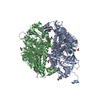

 Mass: 65.409 Da / Num. of mol.: 2 / Source method: obtained synthetically / Formula: Zn
 Mass: 92.094 Da / Num. of mol.: 12 / Source method: obtained synthetically / Formula: C3H8O3
 Mass: 122.143 Da / Num. of mol.: 1 / Source method: obtained synthetically / Formula: C4H12NO3 / Comment: pH buffer*YM
 Mass: 18.015 Da / Num. of mol.: 915 / Source method: isolated from a natural source / Formula: H2O
 Sample preparation
 / Beamline: ID14-2 / Wavelength: 0.933
  Processing