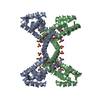
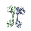
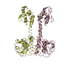
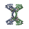
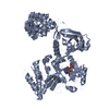
- PDB-2wsh: Structure of bacteriophage T4 EndoII E118A mutant -
+
Open data
ID or keywords:
Loading...
-
Basic information
Entry
Database: PDB / ID: 2wsh
Title
Structure of bacteriophage T4 EndoII E118A mutant
Components
ENDONUCLEASE II
Keywords
HYDROLASE / GIY-YIG / NUCLEASE
Function / homology
Function and homology information
T4 deoxyribonuclease II / degradation of host chromosome by virus / endonuclease activity / symbiont-mediated suppression of host gene expression / hydrolase activity / DNA binding / metal ion binding Similarity search - Function
Mass: 18.015 Da / Num. of mol.: 373 / Source method: isolated from a natural source / Formula: H2O
Compound details
ENGINEERED RESIDUE IN CHAIN A, GLU 126 TO ALA ENGINEERED RESIDUE IN CHAIN B, GLU 126 TO ALA ...ENGINEERED RESIDUE IN CHAIN A, GLU 126 TO ALA ENGINEERED RESIDUE IN CHAIN B, GLU 126 TO ALA ENGINEERED RESIDUE IN CHAIN C, GLU 126 TO ALA ENGINEERED RESIDUE IN CHAIN D, GLU 126 TO ALA
Has protein modification
Y
Sequence details
THE SUBMITTED SEQUENCE IN THIS ENTRY IS 8 AMINO ACIDS SHORTER IN THE N-TERMINUS COMPARED TO OTHER ...THE SUBMITTED SEQUENCE IN THIS ENTRY IS 8 AMINO ACIDS SHORTER IN THE N-TERMINUS COMPARED TO OTHER DATABASE ENTRIES. THE SEQUENCE IS DESCRIBED IN CARLSON ET AL. (1999) , MOL. MICROBIOLOGY, 31(5)., P 1295
-
Experimental details
-
Experiment
Experiment
Method: X-RAY DIFFRACTION / Number of used crystals: 1
-
Sample preparation
Crystal
Density Matthews: 2.6 Å3/Da / Density % sol: 52.9 % Description: THE STARTING MODEL USED FOR MOLECULAR REPLACEMENT AS A PARTIAL MODEL OF ENDOII SOLVED PREVIOUSLY BY SAD.
Crystal grow
Temperature: 293 K / Method: microbatch / pH: 7 Details: CRYSTALS WERE GROWN AT 20 DEGREES, USING THE METHOD MICRO-BATCH UNDER OIL. 2.5 UL PROTEIN SOLUTION (3 MG/ML) WAS MIXED USING VORTEX WITH 1.75 OR 2 UL OF CRYSTALLIZATION SOLUTION CONTAINING ...Details: CRYSTALS WERE GROWN AT 20 DEGREES, USING THE METHOD MICRO-BATCH UNDER OIL. 2.5 UL PROTEIN SOLUTION (3 MG/ML) WAS MIXED USING VORTEX WITH 1.75 OR 2 UL OF CRYSTALLIZATION SOLUTION CONTAINING 30% PEG 3350, 100 MM BIS-TRIS BUFFER PH 7 AND 1% (W/V) N-OCTYL-B-D- GLUCOSIDE AND IMMEDIATELY PLACED UNDER PARAFFIN OIL. THE DROPS WERE IMMEDIATELY STREAK SEEDED AND CRYSTALS GREW IN A FEW HOURS.
In the structure databanks used in Yorodumi, some data are registered as the other names, "COVID-19 virus" and "2019-nCoV". Here are the details of the virus and the list of structure data.
Jan 31, 2019. EMDB accession codes are about to change! (news from PDBe EMDB page)
EMDB accession codes are about to change! (news from PDBe EMDB page)
The allocation of 4 digits for EMDB accession codes will soon come to an end. Whilst these codes will remain in use, new EMDB accession codes will include an additional digit and will expand incrementally as the available range of codes is exhausted. The current 4-digit format prefixed with “EMD-” (i.e. EMD-XXXX) will advance to a 5-digit format (i.e. EMD-XXXXX), and so on. It is currently estimated that the 4-digit codes will be depleted around Spring 2019, at which point the 5-digit format will come into force.
The EM Navigator/Yorodumi systems omit the EMD- prefix.
Related info.:Q: What is EMD? / ID/Accession-code notation in Yorodumi/EM Navigator
Yorodumi is a browser for structure data from EMDB, PDB, SASBDB, etc.
This page is also the successor to EM Navigator detail page, and also detail information page/front-end page for Omokage search.
The word "yorodu" (or yorozu) is an old Japanese word meaning "ten thousand". "mi" (miru) is to see.
Related info.:EMDB / PDB / SASBDB / Comparison of 3 databanks / Yorodumi Search / Aug 31, 2016. New EM Navigator & Yorodumi / Yorodumi Papers / Jmol/JSmol / Function and homology information / Changes in new EM Navigator and Yorodumi
 Movie
Movie Controller


 Open data
 Basic information
 Components
 Keywords
 Function and homology information
 ENTEROBACTERIA PHAGE T4 (virus)
 X-RAY DIFFRACTION /
  Authors
 Citation
 Structure visualization
 Downloads & links
 Other downloads
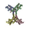
 PDBj
PDBj Assembly
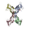


 Mass: 94.971 Da / Num. of mol.: 1 / Source method: obtained synthetically / Formula: PO4
 Mass: 106.120 Da / Num. of mol.: 2 / Source method: obtained synthetically / Formula: C4H10O3
 Mass: 18.015 Da / Num. of mol.: 373 / Source method: isolated from a natural source / Formula: H2O
 Sample preparation
 / Beamline: ID14-1 / Wavelength: 0.933
  Processing