 Movie
Movie Controller

[English] 日本語
 Yorodumi
- PDB-2wqv: Internalin domain of Listeria monocytogenes InlB: rhombohedral cr... -
+ Open data

- Basic information
| Entry | Database: PDB / ID: 2wqv | ||||||
|---|---|---|---|---|---|---|---|
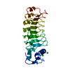
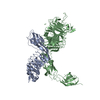
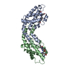
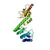
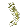
| Title | Internalin domain of Listeria monocytogenes InlB: rhombohedral crystal form | ||||||
 Components Components | INTERNALIN B | ||||||
 Keywords Keywords | CELL INVASION / HGF RECEPTOR LIGAND / LEUCINE-RICH REPEAT / LRR / C-MET LIGAND / VIRULENCE FACTOR | ||||||
| Function / homology |  Function and homology information Function and homology informationpeptidoglycan-based cell wall / InlB-mediated entry of Listeria monocytogenes into host cell / heparin binding / lipid binding / cell surface / extracellular region / metal ion binding / plasma membrane / cytoplasm Similarity search - Function | ||||||
| Biological species |  LISTERIA MONOCYTOGENES (bacteria) LISTERIA MONOCYTOGENES (bacteria) | ||||||
| Method |  X-RAY DIFFRACTION / SYNCHROTRON / MOLECULAR REPLACEMENT / Resolution: 2.8 Å X-RAY DIFFRACTION / SYNCHROTRON / MOLECULAR REPLACEMENT / Resolution: 2.8 Å | ||||||
 Authors Authors | Niemann, H.H. / Heinz, D.W. | ||||||
 Citation Citation | Journal: J.Mol.Biol. / Year: 2010 Title: Ligand-Mediated Dimerization of the met Receptor Tyrosine Kinase by the Bacterial Invasion Protein Inlb. Authors: Ferraris, D.M. / Gherardi, E. / Di, Y. / Heinz, D.W. / Niemann, H.H. | ||||||
| History |
| ||||||
| Remark 700 | SHEET THE SHEET STRUCTURE OF THIS MOLECULE IS BIFURCATED. IN ORDER TO REPRESENT THIS FEATURE IN ... SHEET THE SHEET STRUCTURE OF THIS MOLECULE IS BIFURCATED. IN ORDER TO REPRESENT THIS FEATURE IN THE SHEET RECORDS BELOW, TWO SHEETS ARE DEFINED. |
- Structure visualization
| Structure viewer | Molecule: MolmilJmol/JSmol |
|---|
- Downloads & links
-Download
| PDBx/mmCIF format | 2wqv.cif.gz | 124 KB | Display | PDBx/mmCIF format |
|---|---|---|---|---|
| PDB format | pdb2wqv.ent.gz | 97.4 KB | Display | PDB format |
| PDBx/mmJSON format | 2wqv.json.gz | Tree view | PDBx/mmJSON format | |
| Others |  Other downloads Other downloads |
-Validation report
| Arichive directory | https://data.pdbj.org/pub/pdb/validation_reports/wq/2wqvftp://data.pdbj.org/pub/pdb/validation_reports/wq/2wqv | HTTPS FTP |
|---|
-Related structure data
| Related structure data |  2wquC  2wqwC  2wqxC 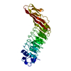 1h6tS S: Starting model for refinement C: citing same article ( |
|---|---|
| Similar structure data |


-Links
 PDBj
PDBj


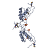
- Assembly
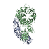
| Deposited unit | 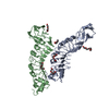
| ||||||||||||||||||||||||||||||||||||||||||||||||||||||||||||||||||||||||||||||||||||||
|---|---|---|---|---|---|---|---|---|---|---|---|---|---|---|---|---|---|---|---|---|---|---|---|---|---|---|---|---|---|---|---|---|---|---|---|---|---|---|---|---|---|---|---|---|---|---|---|---|---|---|---|---|---|---|---|---|---|---|---|---|---|---|---|---|---|---|---|---|---|---|---|---|---|---|---|---|---|---|---|---|---|---|---|---|---|---|---|
| 1 | 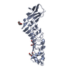
| ||||||||||||||||||||||||||||||||||||||||||||||||||||||||||||||||||||||||||||||||||||||
| 2 | 
| ||||||||||||||||||||||||||||||||||||||||||||||||||||||||||||||||||||||||||||||||||||||
| Unit cell |
| ||||||||||||||||||||||||||||||||||||||||||||||||||||||||||||||||||||||||||||||||||||||
| Noncrystallographic symmetry (NCS) | NCS domain:
NCS domain segments:
NCS ensembles :
NCS oper: (Code: given Matrix: (0.52031, 0.85386, 0.01367), Vector: |
-Components
| #1: Protein | Mass: 32243.818 Da / Num. of mol.: 2 / Fragment: INTERNALIN DOMAIN, RESIDUES 36-321 Source method: isolated from a genetically manipulated source Source: (gene. exp.) LISTERIA MONOCYTOGENES (bacteria) / Strain: EGD-E / Plasmid: PETM30 / Production host: #2: Chemical |   Mass: 194.226 Da / Num. of mol.: 2 / Source method: obtained synthetically / Formula: C8H18O5 / Comment: precipitant*YM Mass: 194.226 Da / Num. of mol.: 2 / Source method: obtained synthetically / Formula: C8H18O5 / Comment: precipitant*YM#3: Chemical | ChemComp-PGE /   Mass: 150.173 Da / Num. of mol.: 4 / Source method: obtained synthetically / Formula: C6H14O4 Mass: 150.173 Da / Num. of mol.: 4 / Source method: obtained synthetically / Formula: C6H14O4#4: Water | ChemComp-HOH / |  Mass: 18.015 Da / Num. of mol.: 31 / Source method: isolated from a natural source / Formula: H2O Mass: 18.015 Da / Num. of mol.: 31 / Source method: isolated from a natural source / Formula: H2OSequence details | RESIDUE 33-35 (GAM) REMAIN AFTER TEV CLEAVAGE OF GST- FUSION PROTEIN | |
|---|
-Experimental details
-Experiment
| Experiment | Method: X-RAY DIFFRACTION / Number of used crystals: 2 |
|---|
- Sample preparation
| Crystal | Density Matthews: 2.97 Å3/Da / Density % sol: 58.6 % / Description: NONE |
|---|---|
| Crystal grow | Temperature: 293 K / Method: vapor diffusion Details: SITTING VAPOR DIFFUSION AT 293 K. 200 NL PROTEIN PLUS 100 NL OF RESERVOIR SOLUTION CONSISTING OF 0.1 M NACL, 0.1 M CHES PH 9.5, 40% PEG300. THE PROTEIN WAS ACTUALLY A COMPLEX OF MET741 WITH ...Details: SITTING VAPOR DIFFUSION AT 293 K. 200 NL PROTEIN PLUS 100 NL OF RESERVOIR SOLUTION CONSISTING OF 0.1 M NACL, 0.1 M CHES PH 9.5, 40% PEG300. THE PROTEIN WAS ACTUALLY A COMPLEX OF MET741 WITH INLB321 AT 5 MG/ML, I.E. INLB321 WAS AT 1.4 MG/ML. CRYSTAL GROWTH TIME: SEVERAL WEEKS TO MONTHS. |
-Data collection
| Diffraction | Mean temperature: 100 K |
|---|---|
| Diffraction source | Source: SYNCHROTRON / Site: ESRF  / Beamline: ID23-2 / Wavelength: 0.873 / Beamline: ID23-2 / Wavelength: 0.873 |
| Detector | Type: MARRESEARCH / Detector: CCD / Date: May 19, 2006 |
| Radiation | Protocol: SINGLE WAVELENGTH / Monochromatic (M) / Laue (L): M / Scattering type: x-ray |
| Radiation wavelength | Wavelength: 0.873 Å / Relative weight: 1 |
| Reflection | Resolution: 2.8→20 Å / Num. obs: 18763 / % possible obs: 99.3 % / Observed criterion σ(I): -3 / Redundancy: 6.96 % / Biso Wilson estimate: 45.1 Å2 / Rmerge(I) obs: 0.16 / Net I/σ(I): 10.98 |
| Reflection shell | Resolution: 2.8→2.87 Å / Redundancy: 7.08 % / Rmerge(I) obs: 0.91 / Mean I/σ(I) obs: 2.58 / % possible all: 100 |
- Processing
| Software |
| ||||||||||||||||||||||||||||||||||||||||||||||||||||||||||||||||||||||||||||||||||||||||||||||||||||||||||||||||||||||||||||||||||||||||||||||||||||||||||||||||||||||||||||||||||||||
|---|---|---|---|---|---|---|---|---|---|---|---|---|---|---|---|---|---|---|---|---|---|---|---|---|---|---|---|---|---|---|---|---|---|---|---|---|---|---|---|---|---|---|---|---|---|---|---|---|---|---|---|---|---|---|---|---|---|---|---|---|---|---|---|---|---|---|---|---|---|---|---|---|---|---|---|---|---|---|---|---|---|---|---|---|---|---|---|---|---|---|---|---|---|---|---|---|---|---|---|---|---|---|---|---|---|---|---|---|---|---|---|---|---|---|---|---|---|---|---|---|---|---|---|---|---|---|---|---|---|---|---|---|---|---|---|---|---|---|---|---|---|---|---|---|---|---|---|---|---|---|---|---|---|---|---|---|---|---|---|---|---|---|---|---|---|---|---|---|---|---|---|---|---|---|---|---|---|---|---|---|---|---|---|
| Refinement | Method to determine structure: MOLECULAR REPLACEMENT Starting model: PDB ENTRY 1H6T Resolution: 2.8→19.822 Å / Cor.coef. Fo:Fc: 0.933 / Cor.coef. Fo:Fc free: 0.917 / SU B: 24.703 / SU ML: 0.233 / TLS residual ADP flag: LIKELY RESIDUAL / Cross valid method: THROUGHOUT / ESU R Free: 0.343 / Stereochemistry target values: MAXIMUM LIKELIHOOD Details: HYDROGENS HAVE BEEN ADDED IN THE RIDING POSITIONS. ATOM RECORD CONTAINS SUM OF TLS AND RESIDUAL B FACTOR
| ||||||||||||||||||||||||||||||||||||||||||||||||||||||||||||||||||||||||||||||||||||||||||||||||||||||||||||||||||||||||||||||||||||||||||||||||||||||||||||||||||||||||||||||||||||||
| Solvent computation | Ion probe radii: 0.8 Å / Shrinkage radii: 0.8 Å / VDW probe radii: 1.2 Å / Solvent model: MASK BULK SOLVENT | ||||||||||||||||||||||||||||||||||||||||||||||||||||||||||||||||||||||||||||||||||||||||||||||||||||||||||||||||||||||||||||||||||||||||||||||||||||||||||||||||||||||||||||||||||||||
| Displacement parameters | Biso mean: 40.35 Å2
| ||||||||||||||||||||||||||||||||||||||||||||||||||||||||||||||||||||||||||||||||||||||||||||||||||||||||||||||||||||||||||||||||||||||||||||||||||||||||||||||||||||||||||||||||||||||
| Refinement step | Cycle: LAST / Resolution: 2.8→19.822 Å
| ||||||||||||||||||||||||||||||||||||||||||||||||||||||||||||||||||||||||||||||||||||||||||||||||||||||||||||||||||||||||||||||||||||||||||||||||||||||||||||||||||||||||||||||||||||||
| Refine LS restraints |
|