 Movie
Movie Controller


+ Open data

- Basic information
| Entry | Database: PDB / ID: 1oto | ||||||
|---|---|---|---|---|---|---|---|
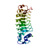
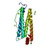
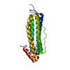
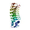
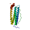
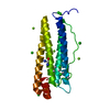
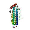
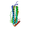
| Title | Calcium-binding mutant of the internalin B LRR domain | ||||||
 Components Components | Internalin B | ||||||
 Keywords Keywords | CELL ADHESION / internalin / InlB / calcium-binding / invasion / Listeria | ||||||
| Function / homology |  Function and homology information Function and homology informationpeptidoglycan-based cell wall / InlB-mediated entry of Listeria monocytogenes into host cell / heparin binding / lipid binding / cell surface / extracellular region / metal ion binding / plasma membrane / cytoplasm Similarity search - Function | ||||||
| Biological species |  Listeria monocytogenes (bacteria) Listeria monocytogenes (bacteria) | ||||||
| Method |  X-RAY DIFFRACTION / MOLECULAR REPLACEMENT / Resolution: 1.96 Å X-RAY DIFFRACTION / MOLECULAR REPLACEMENT / Resolution: 1.96 Å | ||||||
 Authors Authors | Marino, M. / Copp, J. / Dramsi, S. / Chapman, T. / van der Geer, P. / Cossart, P. / Ghosh, P. | ||||||
 Citation Citation | Journal: Biochem.Biophys.Res.Commun. / Year: 2004 Title: Characterization of the calcium-binding sites of Listeria monocytogenes InlB Authors: Marino, M. / Banerjee, M. / Copp, J. / Dramsi, S. / Chapman, T. / Van Der Geer, P. / Cossart, P. / Ghosh, P. | ||||||
| History |
|
- Structure visualization
| Structure viewer | Molecule: MolmilJmol/JSmol |
|---|
- Downloads & links
-Download
| PDBx/mmCIF format | 1oto.cif.gz | 57.9 KB | Display | PDBx/mmCIF format |
|---|---|---|---|---|
| PDB format | pdb1oto.ent.gz | 40.5 KB | Display | PDB format |
| PDBx/mmJSON format | 1oto.json.gz | Tree view | PDBx/mmJSON format | |
| Others |  Other downloads Other downloads |
-Validation report
| Arichive directory | https://data.pdbj.org/pub/pdb/validation_reports/ot/1otoftp://data.pdbj.org/pub/pdb/validation_reports/ot/1oto | HTTPS FTP |
|---|
-Related structure data
| Related structure data |  1otmC  1otnC 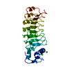 1d0bS S: Starting model for refinement C: citing same article ( |
|---|---|
| Similar structure data |


-Links
 PDBj
PDBj


- Assembly
| Deposited unit | 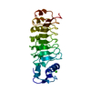
| ||||||||
|---|---|---|---|---|---|---|---|---|---|
| 1 |
| ||||||||
| Unit cell |
|
-Components
| #1: Protein | Mass: 26172.990 Da / Num. of mol.: 1 / Fragment: LRR domain / Mutation: D59A Source method: isolated from a genetically manipulated source Source: (gene. exp.) Listeria monocytogenes (bacteria) / Plasmid: pet28b (Novagen) / Species (production host): Escherichia coli / Production host: |
|---|---|
| #2: Chemical | ChemComp-CA /   Mass: 40.078 Da / Num. of mol.: 1 / Source method: obtained synthetically / Formula: Ca Mass: 40.078 Da / Num. of mol.: 1 / Source method: obtained synthetically / Formula: Ca |
| #3: Water | ChemComp-HOH /  Mass: 18.015 Da / Num. of mol.: 137 / Source method: isolated from a natural source / Formula: H2O Mass: 18.015 Da / Num. of mol.: 137 / Source method: isolated from a natural source / Formula: H2O |
-Experimental details
-Experiment
| Experiment | Method: X-RAY DIFFRACTION / Number of used crystals: 1 |
|---|
- Sample preparation
| Crystal | Density Matthews: 2.09 Å3/Da / Density % sol: 40.77 % | ||||||||||||||||||||||||||||||
|---|---|---|---|---|---|---|---|---|---|---|---|---|---|---|---|---|---|---|---|---|---|---|---|---|---|---|---|---|---|---|---|
| Crystal grow | Temperature: 298 K / Method: vapor diffusion, hanging drop / pH: 6.5 Details: Peg 8000, Mes, Calcium acetate, DTT, pH 6.5, VAPOR DIFFUSION, HANGING DROP, temperature 298K | ||||||||||||||||||||||||||||||
| Crystal grow | *PLUS Method: vapor diffusion / Details: Marino, M., (1999) Mol.Cell, 4, 1063. | ||||||||||||||||||||||||||||||
| Components of the solutions | *PLUS
|
-Data collection
| Diffraction | Mean temperature: 110 K |
|---|---|
| Diffraction source | Source: ROTATING ANODE / Type: RIGAKU / Wavelength: 1.5418 |
| Detector | Type: MARRESEARCH / Detector: IMAGE PLATE / Date: Feb 10, 2000 / Details: Osmic |
| Radiation | Protocol: SINGLE WAVELENGTH / Monochromatic (M) / Laue (L): M / Scattering type: x-ray |
| Radiation wavelength | Wavelength: 1.5418 Å / Relative weight: 1 |
| Reflection | Resolution: 1.96→15 Å / Num. all: 15431 / Num. obs: 15339 / % possible obs: 99.4 % / Observed criterion σ(F): 0 / Observed criterion σ(I): 0 / Redundancy: 4.97 % / Rsym value: 0.053 / Net I/σ(I): 24.23 |
| Reflection shell | Resolution: 1.96→2.03 Å / Mean I/σ(I) obs: 3.18 / Rsym value: 0.493 / % possible all: 99.8 |
| Reflection | *PLUS Lowest resolution: 15 Å / % possible obs: 99 % / Rmerge(I) obs: 0.053 |
| Reflection shell | *PLUS Rmerge(I) obs: 0.493 / Mean I/σ(I) obs: 3.2 |
- Processing
| Software |
| ||||||||||||||||||||
|---|---|---|---|---|---|---|---|---|---|---|---|---|---|---|---|---|---|---|---|---|---|
| Refinement | Method to determine structure: MOLECULAR REPLACEMENT Starting model: pdb entry 1d0b Resolution: 1.96→15 Å / Cross valid method: THROUGHOUT / σ(F): 0 / σ(I): 0 / Stereochemistry target values: Engh & Huber
| ||||||||||||||||||||
| Refinement step | Cycle: LAST / Resolution: 1.96→15 Å
| ||||||||||||||||||||
| Refine LS restraints |
| ||||||||||||||||||||
| Refinement | *PLUS % reflection Rfree: 10 % / Rfactor Rfree: 0.23 / Rfactor Rwork: 0.189 | ||||||||||||||||||||
| Solvent computation | *PLUS | ||||||||||||||||||||
| Displacement parameters | *PLUS | ||||||||||||||||||||
| Refine LS restraints | *PLUS
|
