 Movie
Movie Controller

[English] 日本語
 Yorodumi
- PDB-2wqj: Crystal structure of a truncated variant of the human p73 tetrame... -
+ Open data

- Basic information
| Entry | Database: PDB / ID: 2wqj | ||||||
|---|---|---|---|---|---|---|---|
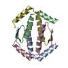
| Title | Crystal structure of a truncated variant of the human p73 tetramerization domain | ||||||
 Components Components | TUMOR PROTEIN P73 | ||||||
 Keywords Keywords | TRANSCRIPTION / P73 / P63 / P53 / TUMOR SUPPRESSION / TRANSCRIPTION FACTOR / TETRAMER / HEXAMER / OLIGOMERIZATION DOMAIN / DNA-BINDING / COOPERATIVITY / CELL-CYCLE CONTROL / TRANSCRIPTION REGULATION / APOPTOSIS / CELL CYCLE / DEVELOPMENT | ||||||
| Function / homology |  Function and homology information Function and homology informationpositive regulation of lung ciliated cell differentiation / cerebrospinal fluid secretion / negative regulation of cardiac muscle cell proliferation / TP53 Regulates Transcription of Death Receptors and Ligands / Activation of PUMA and translocation to mitochondria / positive regulation of intrinsic apoptotic signaling pathway in response to DNA damage by p53 class mediator / Regulation of TP53 Activity through Association with Co-factors / TP53 Regulates Transcription of Caspase Activators and Caspases / digestive tract morphogenesis / negative regulation of neuron differentiation ...positive regulation of lung ciliated cell differentiation / cerebrospinal fluid secretion / negative regulation of cardiac muscle cell proliferation / TP53 Regulates Transcription of Death Receptors and Ligands / Activation of PUMA and translocation to mitochondria / positive regulation of intrinsic apoptotic signaling pathway in response to DNA damage by p53 class mediator / Regulation of TP53 Activity through Association with Co-factors / TP53 Regulates Transcription of Caspase Activators and Caspases / digestive tract morphogenesis / negative regulation of neuron differentiation / TP53 Regulates Transcription of Genes Involved in Cytochrome C Release / TP53 regulates transcription of several additional cell death genes whose specific roles in p53-dependent apoptosis remain uncertain / positive regulation of oligodendrocyte differentiation / positive regulation of cell size / intrinsic apoptotic signaling pathway in response to DNA damage by p53 class mediator / neuron development / mismatch repair / MDM2/MDM4 family protein binding / regulation of mitotic cell cycle / release of cytochrome c from mitochondria / transcription corepressor binding / post-embryonic development / hippocampus development / kidney development / protein tetramerization / promoter-specific chromatin binding / intrinsic apoptotic signaling pathway in response to DNA damage / p53 binding / cell junction / RUNX1 regulates transcription of genes involved in differentiation of HSCs / regulation of gene expression / DNA-binding transcription activator activity, RNA polymerase II-specific / regulation of apoptotic process / DNA-binding transcription factor binding / RNA polymerase II-specific DNA-binding transcription factor binding / negative regulation of neuron apoptotic process / DNA-binding transcription factor activity, RNA polymerase II-specific / positive regulation of MAPK cascade / regulation of cell cycle / transcription cis-regulatory region binding / ciliary basal body / RNA polymerase II cis-regulatory region sequence-specific DNA binding / DNA-binding transcription factor activity / response to xenobiotic stimulus / inflammatory response / negative regulation of cell population proliferation / DNA damage response / centrosome / protein kinase binding / positive regulation of DNA-templated transcription / chromatin / Golgi apparatus / positive regulation of transcription by RNA polymerase II / nucleoplasm / metal ion binding / identical protein binding / nucleus / plasma membrane / cytosol Similarity search - Function | ||||||
| Biological species |  HOMO SAPIENS (human) HOMO SAPIENS (human) | ||||||
| Method |  X-RAY DIFFRACTION / SYNCHROTRON / MAD / Resolution: 2 Å X-RAY DIFFRACTION / SYNCHROTRON / MAD / Resolution: 2 Å | ||||||
 Authors Authors | Joerger, A.C. | ||||||
 Citation Citation | Journal: Proc.Natl.Acad.Sci.USA / Year: 2009 Title: Structural Evolution of P53, P63, and P73: Implication for Heterotetramer Formation. Authors: Joerger, A.C. / Rajagopalan, S. / Natan, E. / Veprintsev, D.B. / Robinson, C.V. / Fersht, A.R. | ||||||
| History |
|
- Structure visualization
| Structure viewer | Molecule: MolmilJmol/JSmol |
|---|
- Downloads & links
-Download
| PDBx/mmCIF format | 2wqj.cif.gz | 179.9 KB | Display | PDBx/mmCIF format |
|---|---|---|---|---|
| PDB format | pdb2wqj.ent.gz | 147.5 KB | Display | PDB format |
| PDBx/mmJSON format | 2wqj.json.gz | Tree view | PDBx/mmJSON format | |
| Others |  Other downloads Other downloads |
-Validation report
| Arichive directory | https://data.pdbj.org/pub/pdb/validation_reports/wq/2wqjftp://data.pdbj.org/pub/pdb/validation_reports/wq/2wqj | HTTPS FTP |
|---|
-Related structure data


-Links
 PDBj
PDBj


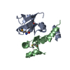
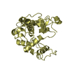
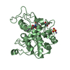
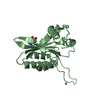
- Assembly
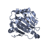
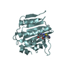
| Deposited unit | 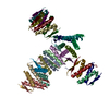
| ||||||||
|---|---|---|---|---|---|---|---|---|---|
| 1 | 
| ||||||||
| 2 | 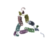
| ||||||||
| 3 | 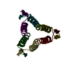
| ||||||||
| 4 | 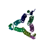
| ||||||||
| 5 | 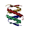
| ||||||||
| Unit cell |
|
-Components
| #1: Protein/peptide | Mass: 4178.738 Da / Num. of mol.: 28 Fragment: TRUNCATED TETRAMERIZATION DOMAIN, RESIDUES 351-383 Source method: isolated from a genetically manipulated source Source: (gene. exp.) HOMO SAPIENS (human) / Production host:  #2: Water | ChemComp-HOH / |  Mass: 18.015 Da / Num. of mol.: 451 / Source method: isolated from a natural source / Formula: H2O Mass: 18.015 Da / Num. of mol.: 451 / Source method: isolated from a natural source / Formula: H2OSequence details | TWO ADDITIONAL | |
|---|
-Experimental details
-Experiment
| Experiment | Method: X-RAY DIFFRACTION / Number of used crystals: 1 |
|---|
- Sample preparation
| Crystal | Density Matthews: 2.9 Å3/Da / Density % sol: 58 % / Description: NONE |
|---|---|
| Crystal grow | Temperature: 290 K / Method: vapor diffusion, sitting drop / pH: 8.5 Details: SITTING DROP VAPOR DIFFUSION AT 17 DEGREE C. PROTEIN SOLUTION: 10 MG/ML IN 20 MM TRIS (PH 8.5), 50 MM NACL. CRYSTALLIZATION BUFFER: 0.1 M TRIS (PH 8.5), 0.9 M AMMONIUM PHOSPHATE. |
-Data collection
| Diffraction | Mean temperature: 100 K |
|---|---|
| Diffraction source | Source: SYNCHROTRON / Site: Diamond  / Beamline: I02 / Wavelength: 0.9796 / Beamline: I02 / Wavelength: 0.9796 |
| Detector | Type: ADSC CCD / Detector: CCD |
| Radiation | Protocol: MAD / Monochromatic (M) / Laue (L): M / Scattering type: x-ray |
| Radiation wavelength | Wavelength: 0.9796 Å / Relative weight: 1 |
| Reflection | Resolution: 2→76.4 Å / Num. obs: 90937 / % possible obs: 99.7 % / Observed criterion σ(I): 3 / Redundancy: 4.8 % / Biso Wilson estimate: 34.31 Å2 / Rmerge(I) obs: 0.06 / Net I/σ(I): 16.2 |
| Reflection shell | Resolution: 2→2.11 Å / Redundancy: 3.7 % / Rmerge(I) obs: 0.37 / Mean I/σ(I) obs: 3.7 / % possible all: 99.3 |
- Processing
| Software | Name: PHENIX / Version: (PHENIX.REFINE) / Classification: refinement | |||||||||||||||||||||||||||||||||||||||||||||||||||||||||||||||||||||||||||||||||||||||||||||||||||||||||||||||||||||||||||||||||||||||||||||||||||||||||||||||||||||||||||||||||||||||||||||||||||||||||||||||||||||||||
|---|---|---|---|---|---|---|---|---|---|---|---|---|---|---|---|---|---|---|---|---|---|---|---|---|---|---|---|---|---|---|---|---|---|---|---|---|---|---|---|---|---|---|---|---|---|---|---|---|---|---|---|---|---|---|---|---|---|---|---|---|---|---|---|---|---|---|---|---|---|---|---|---|---|---|---|---|---|---|---|---|---|---|---|---|---|---|---|---|---|---|---|---|---|---|---|---|---|---|---|---|---|---|---|---|---|---|---|---|---|---|---|---|---|---|---|---|---|---|---|---|---|---|---|---|---|---|---|---|---|---|---|---|---|---|---|---|---|---|---|---|---|---|---|---|---|---|---|---|---|---|---|---|---|---|---|---|---|---|---|---|---|---|---|---|---|---|---|---|---|---|---|---|---|---|---|---|---|---|---|---|---|---|---|---|---|---|---|---|---|---|---|---|---|---|---|---|---|---|---|---|---|---|---|---|---|---|---|---|---|---|---|---|---|---|---|---|---|---|
| Refinement | Method to determine structure: MAD Starting model: NONE Resolution: 2→24.96 Å / SU ML: 0.29 / σ(F): 1.08 / Phase error: 25.49 / Stereochemistry target values: ML Details: THIS ENTRY REPORTS THE STRUCTURE OF A TRUNCATED FORM OF THE P73 TETRAMERIZATION DOMAIN (RESIDUES 351-383)THAT LACKS A C-TERMINAL HELIX THAT IS ESSENTIAL FOR STABILIZING THE OVERALL ...Details: THIS ENTRY REPORTS THE STRUCTURE OF A TRUNCATED FORM OF THE P73 TETRAMERIZATION DOMAIN (RESIDUES 351-383)THAT LACKS A C-TERMINAL HELIX THAT IS ESSENTIAL FOR STABILIZING THE OVERALL ARCHITECTURE OF THE P73 TETRAMER. SEE PDB ENTRY 2WQI WITH THE STRUCTURE OF P73 RESIDUES 351-399 FOR COMPARISON.
| |||||||||||||||||||||||||||||||||||||||||||||||||||||||||||||||||||||||||||||||||||||||||||||||||||||||||||||||||||||||||||||||||||||||||||||||||||||||||||||||||||||||||||||||||||||||||||||||||||||||||||||||||||||||||
| Solvent computation | Shrinkage radii: 0.9 Å / VDW probe radii: 1.11 Å / Solvent model: FLAT BULK SOLVENT MODEL / Bsol: 50.14 Å2 / ksol: 0.349 e/Å3 | |||||||||||||||||||||||||||||||||||||||||||||||||||||||||||||||||||||||||||||||||||||||||||||||||||||||||||||||||||||||||||||||||||||||||||||||||||||||||||||||||||||||||||||||||||||||||||||||||||||||||||||||||||||||||
| Displacement parameters | Biso mean: 39.6 Å2
| |||||||||||||||||||||||||||||||||||||||||||||||||||||||||||||||||||||||||||||||||||||||||||||||||||||||||||||||||||||||||||||||||||||||||||||||||||||||||||||||||||||||||||||||||||||||||||||||||||||||||||||||||||||||||
| Refinement step | Cycle: LAST / Resolution: 2→24.96 Å
| |||||||||||||||||||||||||||||||||||||||||||||||||||||||||||||||||||||||||||||||||||||||||||||||||||||||||||||||||||||||||||||||||||||||||||||||||||||||||||||||||||||||||||||||||||||||||||||||||||||||||||||||||||||||||
| Refine LS restraints |
| |||||||||||||||||||||||||||||||||||||||||||||||||||||||||||||||||||||||||||||||||||||||||||||||||||||||||||||||||||||||||||||||||||||||||||||||||||||||||||||||||||||||||||||||||||||||||||||||||||||||||||||||||||||||||
| LS refinement shell |
|
