Mass: 18.015 Da / Num. of mol.: 382 / Source method: isolated from a natural source / Formula: H2O
-
Details
Compound details
ENGINEERED RESIDUE IN CHAIN A, SER 51 TO PRO ENGINEERED RESIDUE IN CHAIN A, MET 111 TO VAL ...ENGINEERED RESIDUE IN CHAIN A, SER 51 TO PRO ENGINEERED RESIDUE IN CHAIN A, MET 111 TO VAL ENGINEERED RESIDUE IN CHAIN A, GLY 236 TO GLU ENGINEERED RESIDUE IN CHAIN A, TRP 331 TO PHE ENGINEERED RESIDUE IN CHAIN A, ARG 371 TO LYS ENGINEERED RESIDUE IN CHAIN A, GLN 447 TO THR ENGINEERED RESIDUE IN CHAIN A, PRO 504 TO ILE ENGINEERED RESIDUE IN CHAIN A, VAL 535 TO ALA ENGINEERED RESIDUE IN CHAIN A, ASN 576 TO ASP
Has protein modification
Y
-
Experimental details
-
Experiment
Experiment
Method: X-RAY DIFFRACTION / Number of used crystals: 1
-
Sample preparation
Crystal
Density Matthews: 3.14 Å3/Da / Density % sol: 60.52 % / Description: NONE
Crystal grow
pH: 5 Details: 10% PEG6K, 0.1 M CACL2, 5% GLYCEROL, 10 MM GLCNAC, 50 M ACETATE PH 5.
Resolution: 2.19→76.7 Å / Cor.coef. Fo:Fc: 0.931 / Cor.coef. Fo:Fc free: 0.887 / SU B: 4.058 / SU ML: 0.105 / Cross valid method: THROUGHOUT / ESU R: 0.221 / ESU R Free: 0.204 / Stereochemistry target values: MAXIMUM LIKELIHOOD Details: HYDROGENS HAVE BEEN ADDED IN THE RIDING POSITIONS. U VALUES REFINED INDIVIDUALLY.
Rfactor
Num. reflection
% reflection
Selection details
Rfree
0.24175
2174
5.1 %
RANDOM
Rwork
0.18212
-
-
-
obs
0.18515
40873
85.33 %
-
Solvent computation
Ion probe radii: 0.8 Å / Shrinkage radii: 0.8 Å / VDW probe radii: 1.2 Å / Solvent model: MASK
 Movie
Movie Controller

 Yorodumi
 Open data
 Basic information
 Components
 Keywords
 Function and homology information
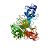
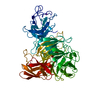
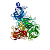
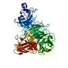
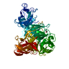
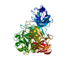
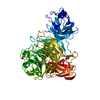
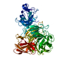
 GIBBERELLA ZEAE (fungus)
 X-RAY DIFFRACTION /
  Authors
 Citation
 Structure visualization
 Downloads & links
 Other downloads
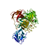


 PDBj
PDBj


 Assembly


 Mass: 63.546 Da / Num. of mol.: 1 / Source method: obtained synthetically / Formula: Cu
 Mass: 40.078 Da / Num. of mol.: 1 / Source method: obtained synthetically / Formula: Ca
 Mass: 60.052 Da / Num. of mol.: 2 / Source method: obtained synthetically / Formula: C2H4O2
 Mass: 62.068 Da / Num. of mol.: 9 / Source method: obtained synthetically / Formula: C2H6O2
 Mass: 92.094 Da / Num. of mol.: 3 / Source method: obtained synthetically / Formula: C3H8O3
 Sample preparation
 / Beamline: ID14-1 / Wavelength: 0.934
  Processing