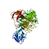
 Movie
Movie Controller


+ Open data

- Basic information
| Entry | Database: PDB / ID: 2eib | ||||||
|---|---|---|---|---|---|---|---|
| Title | Crystal Structure of Galactose Oxidase, W290H mutant | ||||||
 Components Components | Galactose oxidase | ||||||
 Keywords Keywords | OXIDOREDUCTASE / Galactose Oxidase mutant | ||||||
| Function / homology |  Function and homology information Function and homology informationgalactose oxidase / galactose oxidase activity / extracellular region / metal ion binding Similarity search - Function | ||||||
| Biological species |  Gibberella zeae (fungus) Gibberella zeae (fungus) | ||||||
| Method |  X-RAY DIFFRACTION / FOURIER SYNTHESIS / Resolution: 2.1 Å X-RAY DIFFRACTION / FOURIER SYNTHESIS / Resolution: 2.1 Å | ||||||
 Authors Authors | Phillips, S.E. / McPherson, M.J. / Knowles, P.F. / Wilmot, C. | ||||||
 Citation Citation | Journal: Biochemistry / Year: 2007 Title: The Stacking Tryptophan of Galactose Oxidase: A Second-Coordination Sphere Residue that Has Profound Effects on Tyrosyl Radical Behavior and Enzyme Catalysis Authors: Rogers, M.S. / Tyler, E.M. / Akyumani, N. / Kurtis, C.R. / Spooner, R.K. / Deacon, S.E. / Tamber, S. / Firbank, S.J. / Mahmoud, K. / Knowles, P.F. / Phillips, S.E. / McPherson, M.J. / Dooley, D.M. #1: Journal: J.Biol.Chem. / Year: 1994 Title: Structure and mechanism of galactose oxidase. The free radical site Authors: Baron, A.J. / Stevens, C. / Wilmot, C. / Seneviratne, K.D. / Blakeley, V. / Dooley, D.M. / Phillips, S.E. / Knowles, P.F. / McPherson, M.J. | ||||||
| History |
|
- Structure visualization
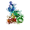
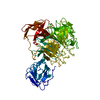
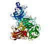
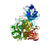
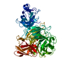
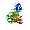
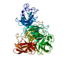
| Structure viewer | Molecule: MolmilJmol/JSmol |
|---|
- Downloads & links
-Download
| PDBx/mmCIF format | 2eib.cif.gz | 143.1 KB | Display | PDBx/mmCIF format |
|---|---|---|---|---|
| PDB format | pdb2eib.ent.gz | 108 KB | Display | PDB format |
| PDBx/mmJSON format | 2eib.json.gz | Tree view | PDBx/mmJSON format | |
| Others |  Other downloads Other downloads |
-Validation report
| Arichive directory | https://data.pdbj.org/pub/pdb/validation_reports/ei/2eibftp://data.pdbj.org/pub/pdb/validation_reports/ei/2eib | HTTPS FTP |
|---|
-Related structure data
| Related structure data |  2eicC 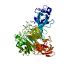 2eidC  2eieC  1gogS C: citing same article ( S: Starting model for refinement |
|---|---|
| Similar structure data |


-Links
 PDBj
PDBj

- Assembly
| Deposited unit | 
| ||||||||
|---|---|---|---|---|---|---|---|---|---|
| 1 |
| ||||||||
| Unit cell |
| ||||||||
| Details | Protein is a monomer in the asymmetric unit |
-Components
-Protein , 1 types, 1 molecules A
| #1: Protein | Mass: 68530.531 Da / Num. of mol.: 1 / Fragment: residues 1-639 / Mutation: W290H Source method: isolated from a genetically manipulated source Source: (gene. exp.) Gibberella zeae (fungus) / Gene: go / Plasmid: pGOF1 / Production host: References: UniProt: Q01745, UniProt: P0CS93*PLUS, galactose oxidase |
|---|
-Non-polymers , 5 types, 332 molecules 


| #2: Chemical | ChemComp-CU /  Mass: 63.546 Da / Num. of mol.: 1 / Source method: obtained synthetically / Formula: Cu Mass: 63.546 Da / Num. of mol.: 1 / Source method: obtained synthetically / Formula: Cu | ||
|---|---|---|---|
| #3: Chemical | ChemComp-NA /  Mass: 22.990 Da / Num. of mol.: 1 / Source method: obtained synthetically / Formula: Na Mass: 22.990 Da / Num. of mol.: 1 / Source method: obtained synthetically / Formula: Na | ||
| #4: Chemical | ChemComp-SO4 /  Mass: 96.063 Da / Num. of mol.: 1 / Source method: obtained synthetically / Formula: SO4 Mass: 96.063 Da / Num. of mol.: 1 / Source method: obtained synthetically / Formula: SO4 | ||
| #5: Chemical |  Mass: 59.044 Da / Num. of mol.: 3 / Source method: obtained synthetically / Formula: C2H3O2 Mass: 59.044 Da / Num. of mol.: 3 / Source method: obtained synthetically / Formula: C2H3O2#6: Water | ChemComp-HOH / | Mass: 18.015 Da / Num. of mol.: 326 / Source method: isolated from a natural source / Formula: H2O |
-Details
| Has protein modification | Y |
|---|
-Experimental details
-Experiment
| Experiment | Method: X-RAY DIFFRACTION / Number of used crystals: 1 |
|---|
- Sample preparation
| Crystal | Density Matthews: 4.9 Å3/Da / Density % sol: 74.9 % |
|---|---|
| Crystal grow | Temperature: 298 K / Method: vapor diffusion, sitting drop / pH: 4.5 Details: 1-2M Amonium Sulphate, 0.1M sodium acetate pH 4-5, pH 4.5, VAPOR DIFFUSION, SITTING DROP, temperature 298K |
-Data collection
| Diffraction | Mean temperature: 298 K |
|---|---|
| Diffraction source | Source: ROTATING ANODE / Type: RIGAKU RU200 / Wavelength: 1.5418 Å |
| Detector | Type: SIEMENS-NICOLET X100 / Detector: AREA DETECTOR |
| Radiation | Monochromator: Graphite / Protocol: SINGLE WAVELENGTH / Monochromatic (M) / Laue (L): M / Scattering type: x-ray |
| Radiation wavelength | Wavelength: 1.5418 Å / Relative weight: 1 |
| Reflection | Resolution: 2.1→30 Å / Rsym value: 0.046 |
- Processing
| Software | Name: PROLSQ / Classification: refinement | ||||||||||||
|---|---|---|---|---|---|---|---|---|---|---|---|---|---|
| Refinement | Method to determine structure: FOURIER SYNTHESIS Starting model: 1GOG Resolution: 2.1→10 Å / Rfactor Rwork: 0.157 / σ(F): 0 / σ(I): 0 / Stereochemistry target values: Engh & Huber | ||||||||||||
| Displacement parameters | Biso mean: 24.376 Å2 | ||||||||||||
| Refinement step | Cycle: LAST / Resolution: 2.1→10 Å
| ||||||||||||
| Refine LS restraints | Type: p_bond_d / Dev ideal: 0.017 |
