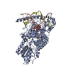
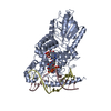
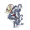
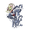
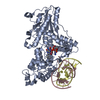
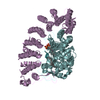
Resolution: 2→40.468 Å / SU ML: 0.43 / σ(F): 1.35 / Phase error: 19.97 / Stereochemistry target values: ML Details: RESIDUES DT 8 AND DC 9 HAVE FORMED A 6-4 PHOTOPRODUCT BY THE COVALENT BONDING OF ATOM C6 OF TDY TO C4 OF Z.
Rfactor
Num. reflection
% reflection
Rfree
0.2089
2451
5.1 %
Rwork
0.1778
-
-
obs
0.1794
48536
99.02 %
Solvent computation
Shrinkage radii: 0.9 Å / VDW probe radii: 1.11 Å / Solvent model: FLAT BULK SOLVENT MODEL / Bsol: 40 Å2 / ksol: 0.351 e/Å3
Displacement parameters
Biso mean: 28.4 Å2
Baniso -1
Baniso -2
Baniso -3
1-
0.8264 Å2
0 Å2
0 Å2
2-
-
0.8688 Å2
0 Å2
3-
-
-
-3.1512 Å2
Refinement step
Cycle: LAST / Resolution: 2→40.468 Å
Protein
Nucleic acid
Ligand
Solvent
Total
Num. atoms
4162
609
53
403
5227
Refine LS restraints
Refine-ID
Type
Dev ideal
Number
X-RAY DIFFRACTION
f_bond_d
0.011
5058
X-RAY DIFFRACTION
f_angle_d
1.377
7002
X-RAY DIFFRACTION
f_dihedral_angle_d
19.071
1938
X-RAY DIFFRACTION
f_chiral_restr
0.103
727
X-RAY DIFFRACTION
f_plane_restr
0.007
790
LS refinement shell
Resolution (Å)
Rfactor Rfree
Num. reflection Rfree
Rfactor Rwork
Num. reflection Rwork
Refine-ID
% reflection obs (%)
1.9955-2.0338
0.3023
135
0.2361
2307
X-RAY DIFFRACTION
90
2.0338-2.0754
0.2956
117
0.2222
2546
X-RAY DIFFRACTION
99
2.0754-2.1205
0.2568
136
0.1921
2519
X-RAY DIFFRACTION
100
2.1205-2.1698
0.2356
121
0.1791
2574
X-RAY DIFFRACTION
100
2.1698-2.2241
0.21
147
0.1781
2517
X-RAY DIFFRACTION
100
2.2241-2.2842
0.2081
149
0.1822
2554
X-RAY DIFFRACTION
100
2.2842-2.3514
0.256
144
0.1779
2530
X-RAY DIFFRACTION
100
2.3514-2.4273
0.2413
123
0.1755
2558
X-RAY DIFFRACTION
100
2.4273-2.514
0.2368
155
0.1735
2535
X-RAY DIFFRACTION
100
2.514-2.6147
0.2121
156
0.1792
2559
X-RAY DIFFRACTION
100
2.6147-2.7336
0.2755
124
0.1876
2565
X-RAY DIFFRACTION
100
2.7336-2.8777
0.2532
134
0.2007
2584
X-RAY DIFFRACTION
100
2.8777-3.058
0.2383
128
0.1971
2576
X-RAY DIFFRACTION
100
3.058-3.294
0.2099
130
0.1859
2597
X-RAY DIFFRACTION
100
3.294-3.6253
0.1874
134
0.1685
2600
X-RAY DIFFRACTION
100
3.6253-4.1494
0.1655
137
0.1436
2623
X-RAY DIFFRACTION
100
4.1494-5.226
0.149
141
0.1408
2616
X-RAY DIFFRACTION
99
5.226-40.4761
0.1615
140
0.1623
2725
X-RAY DIFFRACTION
98
+
About Yorodumi
-
News
-
Feb 9, 2022. New format data for meta-information of EMDB entries
New format data for meta-information of EMDB entries
Version 3 of the EMDB header file is now the official format.
The previous official version 1.9 will be removed from the archive.
In the structure databanks used in Yorodumi, some data are registered as the other names, "COVID-19 virus" and "2019-nCoV". Here are the details of the virus and the list of structure data.
Jan 31, 2019. EMDB accession codes are about to change! (news from PDBe EMDB page)
EMDB accession codes are about to change! (news from PDBe EMDB page)
The allocation of 4 digits for EMDB accession codes will soon come to an end. Whilst these codes will remain in use, new EMDB accession codes will include an additional digit and will expand incrementally as the available range of codes is exhausted. The current 4-digit format prefixed with “EMD-” (i.e. EMD-XXXX) will advance to a 5-digit format (i.e. EMD-XXXXX), and so on. It is currently estimated that the 4-digit codes will be depleted around Spring 2019, at which point the 5-digit format will come into force.
The EM Navigator/Yorodumi systems omit the EMD- prefix.
Related info.:Q: What is EMD? / ID/Accession-code notation in Yorodumi/EM Navigator
Yorodumi is a browser for structure data from EMDB, PDB, SASBDB, etc.
This page is also the successor to EM Navigator detail page, and also detail information page/front-end page for Omokage search.
The word "yorodu" (or yorozu) is an old Japanese word meaning "ten thousand". "mi" (miru) is to see.
Related info.:EMDB / PDB / SASBDB / Comparison of 3 databanks / Yorodumi Search / Aug 31, 2016. New EM Navigator & Yorodumi / Yorodumi Papers / Jmol/JSmol / Function and homology information / Changes in new EM Navigator and Yorodumi
 Movie
Movie Controller

 Yorodumi
 Open data
 Basic information
 Components
 Keywords
 Function and homology information
 X-RAY DIFFRACTION /
  Authors
 Citation
 Structure visualization
 Downloads & links
 Other downloads


 PDBj
PDBj


 Assembly


 Mass: 785.550 Da / Num. of mol.: 1 / Source method: obtained synthetically / Formula: C27H33N9O15P2 / Comment: FAD*YM
 Mass: 18.015 Da / Num. of mol.: 403 / Source method: isolated from a natural source / Formula: H2O
 Sample preparation
 / Beamline: X10SA / Wavelength: 1
  Processing