 Movie
Movie Controller

[English] 日本語
 Yorodumi
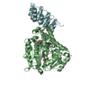
- PDB-2wb2: Drosophila Melanogaster (6-4) Photolyase Bound To double stranded... -
+ Open data

- Basic information
| Entry | Database: PDB / ID: 2wb2 | |||||||||
|---|---|---|---|---|---|---|---|---|---|---|
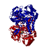
| Title | Drosophila Melanogaster (6-4) Photolyase Bound To double stranded Dna containing a T(6-4)C Photolesion | |||||||||
 Components Components |
| |||||||||
 Keywords Keywords | LYASE/DNA / LYASE-DNA COMPLEX / PHOTOLESION / DNA PHOTOLYASE / LYASE | |||||||||
| Function / homology |  Function and homology information Function and homology information | |||||||||
| Biological species |  synthetic construct (others) | |||||||||
| Method |  X-RAY DIFFRACTION / SYNCHROTRON / MOLECULAR REPLACEMENT / Resolution: 2.95 Å X-RAY DIFFRACTION / SYNCHROTRON / MOLECULAR REPLACEMENT / Resolution: 2.95 Å | |||||||||
 Authors Authors | Glas, A.F. / Schneider, S. / Maul, M.J. / Hennecke, U. / Carell, T. | |||||||||
 Citation Citation | Journal: Chemistry / Year: 2009 Title: Crystal Structure of the T(6-4)C Lesion in Complex with a (6-4) DNA Photolyase and Repair of Uv- Induced (6-4) and Dewar Photolesions. Authors: Glas, A.F. / Schneider, S. / Maul, M.J. / Hennecke, U. / Carell, T. #1: Journal: Angew.Chem.Int.Ed.Engl. / Year: 2008Title: Crystal Structure and Mechanism of a DNA (6-4) Photolyase. Authors: Maul, M.J. / Barends, T.R.M. / Glas, A.F. / Cryle, M.J. / Domratcheva, T. / Schneider, S. / Schlichting, I. / Carell, T. | |||||||||
| History |
|
- Structure visualization
| Structure viewer | Molecule: MolmilJmol/JSmol |
|---|
- Downloads & links
-Download
| PDBx/mmCIF format | 2wb2.cif.gz | 142.2 KB | Display | PDBx/mmCIF format |
|---|---|---|---|---|
| PDB format | pdb2wb2.ent.gz | 105.2 KB | Display | PDB format |
| PDBx/mmJSON format | 2wb2.json.gz | Tree view | PDBx/mmJSON format | |
| Others |  Other downloads Other downloads |
-Validation report
| Arichive directory | https://data.pdbj.org/pub/pdb/validation_reports/wb/2wb2ftp://data.pdbj.org/pub/pdb/validation_reports/wb/2wb2 | HTTPS FTP |
|---|
-Related structure data
| Related structure data |  3cvuS S: Starting model for refinement |
|---|---|
| Similar structure data |


-Links
 PDBj
PDBj


- Assembly
| Deposited unit | 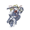
| ||||||||
|---|---|---|---|---|---|---|---|---|---|
| 1 |
| ||||||||
| Unit cell |
|
-Components
| #1: Protein | Mass: 62910.137 Da / Num. of mol.: 1 / Fragment: RESIDUES 1-520 Source method: isolated from a genetically manipulated source Source: (gene. exp.)  References: UniProt: Q8SXK5, deoxyribodipyrimidine photo-lyase |
|---|---|
| #2: DNA chain | Mass: 4637.026 Da / Num. of mol.: 1 / Source method: obtained synthetically / Details: 6-4 LINK BETWEEN T 8 AND C 8 (SEE REMARK 600) / Source: (synth.) synthetic construct (others) |
| #3: DNA chain | Mass: 4545.948 Da / Num. of mol.: 1 / Source method: obtained synthetically / Source: (synth.) synthetic construct (others) |
| #4: Chemical | ChemComp-FAD /   Mass: 785.550 Da / Num. of mol.: 1 / Source method: obtained synthetically / Formula: C27H33N9O15P2 / Comment: FAD*YM Mass: 785.550 Da / Num. of mol.: 1 / Source method: obtained synthetically / Formula: C27H33N9O15P2 / Comment: FAD*YM |
| Nonpolymer details | CHAIN C HETEROGENS 64P AND Z: RESIDUES DT 8 AND DC 9 HAVE FORMED A PHOTOPRODUCT BY THE COVALENT ...CHAIN C HETEROGENS |
| Sequence details | CONSTRUCT CONTAINS RESIDUES 1-520 |
-Experimental details
-Experiment
| Experiment | Method: X-RAY DIFFRACTION / Number of used crystals: 1 |
|---|
- Sample preparation
| Crystal | Density Matthews: 2.48 Å3/Da / Density % sol: 50 % / Description: NONE |
|---|---|
| Crystal grow | Temperature: 291 K / Method: vapor diffusion, hanging drop Details: PROTEIN WAS MIXED WITH DNA IN A 1:1.1 MOLAR RATIO WITH A FINAL CONCENTRATION OF 8.5MG/ML PROTEIN. 0.1M HEPES PH 7, 15-20% POLYETHYLENGLYCOL 4000, HANGING-DROP VAPOUR DIFFUSION |
-Data collection
| Diffraction | Mean temperature: 100 K |
|---|---|
| Diffraction source | Source: SYNCHROTRON / Site: SLS  / Beamline: X06SA / Wavelength: 1.076 / Beamline: X06SA / Wavelength: 1.076 |
| Detector | Type: MARRESEARCH / Detector: CCD / Date: Jul 4, 2008 |
| Radiation | Protocol: SINGLE WAVELENGTH / Monochromatic (M) / Laue (L): M / Scattering type: x-ray |
| Radiation wavelength | Wavelength: 1.076 Å / Relative weight: 1 |
| Reflection | Resolution: 2.95→45.8 Å / Num. obs: 15653 / % possible obs: 99.9 % / Observed criterion σ(I): 2 / Redundancy: 5.3 % / Rmerge(I) obs: 0.07 / Net I/σ(I): 13.1 |
| Reflection shell | Resolution: 2.95→3.11 Å / Redundancy: 5.6 % / Rmerge(I) obs: 0.41 / Mean I/σ(I) obs: 3.1 / % possible all: 100 |
- Processing
| Software |
| ||||||||||||||||||||||||||||||||||||||||||||||||||||||||||||||||||||||||||||||||||||||||||||||||||||||||||||||||||||||||||||||||||||||||||||||||||||||||||||||||||||||||||||||||||||||
|---|---|---|---|---|---|---|---|---|---|---|---|---|---|---|---|---|---|---|---|---|---|---|---|---|---|---|---|---|---|---|---|---|---|---|---|---|---|---|---|---|---|---|---|---|---|---|---|---|---|---|---|---|---|---|---|---|---|---|---|---|---|---|---|---|---|---|---|---|---|---|---|---|---|---|---|---|---|---|---|---|---|---|---|---|---|---|---|---|---|---|---|---|---|---|---|---|---|---|---|---|---|---|---|---|---|---|---|---|---|---|---|---|---|---|---|---|---|---|---|---|---|---|---|---|---|---|---|---|---|---|---|---|---|---|---|---|---|---|---|---|---|---|---|---|---|---|---|---|---|---|---|---|---|---|---|---|---|---|---|---|---|---|---|---|---|---|---|---|---|---|---|---|---|---|---|---|---|---|---|---|---|---|---|
| Refinement | Method to determine structure: MOLECULAR REPLACEMENT Starting model: PDB ENTRY 3CVU Resolution: 2.95→45 Å / Cor.coef. Fo:Fc: 0.931 / Cor.coef. Fo:Fc free: 0.912 / SU B: 38.342 / SU ML: 0.337 / TLS residual ADP flag: LIKELY RESIDUAL / Cross valid method: THROUGHOUT / ESU R Free: 0.415 / Stereochemistry target values: MAXIMUM LIKELIHOOD Details: HYDROGENS HAVE BEEN ADDED IN THE RIDING POSITIONS. U VALUES RESIDUAL ONLY
| ||||||||||||||||||||||||||||||||||||||||||||||||||||||||||||||||||||||||||||||||||||||||||||||||||||||||||||||||||||||||||||||||||||||||||||||||||||||||||||||||||||||||||||||||||||||
| Solvent computation | Ion probe radii: 0.8 Å / Shrinkage radii: 0.8 Å / VDW probe radii: 1.2 Å / Solvent model: MASK | ||||||||||||||||||||||||||||||||||||||||||||||||||||||||||||||||||||||||||||||||||||||||||||||||||||||||||||||||||||||||||||||||||||||||||||||||||||||||||||||||||||||||||||||||||||||
| Displacement parameters | Biso mean: 63.264 Å2
| ||||||||||||||||||||||||||||||||||||||||||||||||||||||||||||||||||||||||||||||||||||||||||||||||||||||||||||||||||||||||||||||||||||||||||||||||||||||||||||||||||||||||||||||||||||||
| Refinement step | Cycle: LAST / Resolution: 2.95→45 Å
| ||||||||||||||||||||||||||||||||||||||||||||||||||||||||||||||||||||||||||||||||||||||||||||||||||||||||||||||||||||||||||||||||||||||||||||||||||||||||||||||||||||||||||||||||||||||
| Refine LS restraints |
|