 Movie
Movie Controller

[English] 日本語
 Yorodumi
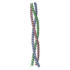
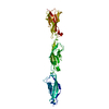
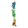
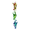
- PDB-2wpq: Salmonella enterica SadA 479-519 fused to GCN4 adaptors (SadAK3, ... -
+ Open data

- Basic information
| Entry | Database: PDB / ID: 2wpq | ||||||
|---|---|---|---|---|---|---|---|
| Title | Salmonella enterica SadA 479-519 fused to GCN4 adaptors (SadAK3, in- register fusion) | ||||||
 Components Components | TRIMERIC AUTOTRANSPORTER ADHESIN FRAGMENT | ||||||
 Keywords Keywords | MEMBRANE PROTEIN / HYDROPHOBIC CORE / ION COORDINATION / PROTEIN EXPORT / TRIMERIC AUTOTRANSPORTER ADHESIN / TAA / POLAR CORE RESIDUES | ||||||
| Function / homology |  Function and homology information Function and homology information | ||||||
| Biological species |  SALMONELLA ENTERICA SUBSP. ENTERICA SEROVAR TYPHIMURIUM (bacteria) SALMONELLA ENTERICA SUBSP. ENTERICA SEROVAR TYPHIMURIUM (bacteria) | ||||||
| Method |  X-RAY DIFFRACTION / SYNCHROTRON / MOLECULAR REPLACEMENT / Resolution: 1.85 Å X-RAY DIFFRACTION / SYNCHROTRON / MOLECULAR REPLACEMENT / Resolution: 1.85 Å | ||||||
 Authors Authors | Hartmann, M.D. / Hernandez Alvarez, B. / Albrecht, R. / Zeth, K. / Lupas, A.N. | ||||||
 Citation Citation | Journal: Proc.Natl.Acad.Sci.USA / Year: 2009 Title: A Coiled-Coil Motif that Sequesters Ions to the Hydrophobic Core. Authors: Hartmann, M.D. / Ridderbusch, O. / Zeth, K. / Albrecht, R. / Testa, O. / Woolfson, D.N. / Sauer, G. / Dunin-Horkawicz, S. / Lupas, A.N. / Alvarez, B.H. #1: Journal: Protein Eng.Des.Sel. / Year: 2008Title: A New Expression System for Protein Crystallization Using Trimeric Coiled-Coil Adaptors. Authors: Hernandez Alvarez, B. / Hartmann, M.D. / Albrecht, R. / Lupas, A.N. / Zeth, K. / Linke, D. | ||||||
| History |
|
- Structure visualization
| Structure viewer | Molecule: MolmilJmol/JSmol |
|---|
- Downloads & links
-Download
| PDBx/mmCIF format | 2wpq.cif.gz | 74.9 KB | Display | PDBx/mmCIF format |
|---|---|---|---|---|
| PDB format | pdb2wpq.ent.gz | 57.3 KB | Display | PDB format |
| PDBx/mmJSON format | 2wpq.json.gz | Tree view | PDBx/mmJSON format | |
| Others |  Other downloads Other downloads |
-Validation report
| Arichive directory | https://data.pdbj.org/pub/pdb/validation_reports/wp/2wpqftp://data.pdbj.org/pub/pdb/validation_reports/wp/2wpq | HTTPS FTP |
|---|
-Related structure data
| Related structure data |  2wprC  2wpsC  2wpyC  2wpzC  2wq0C 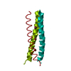 2wq1C 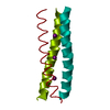 2wq2C  2wq3C  1gcmS C: citing same article ( S: Starting model for refinement |
|---|---|
| Similar structure data |


-Links
 PDBj
PDBj- Assembly
| Deposited unit | 
| ||||||||||||||||||||||||
|---|---|---|---|---|---|---|---|---|---|---|---|---|---|---|---|---|---|---|---|---|---|---|---|---|---|
| 1 |
| ||||||||||||||||||||||||
| Unit cell |
| ||||||||||||||||||||||||
| Noncrystallographic symmetry (NCS) | NCS domain:
NCS domain segments: Component-ID: 1 / Ens-ID: 1 / Beg auth comp-ID: MET / Beg label comp-ID: MET / End auth comp-ID: ILE / End label comp-ID: ILE / Refine code: 6 / Auth seq-ID: 450 - 548 / Label seq-ID: 1 - 99
|
-Components
| #1: Protein | Mass: 11532.017 Da / Num. of mol.: 3 / Fragment: RESIDUES 479-519 FUSED TO GCN4 ADAPTORS Source method: isolated from a genetically manipulated source Details: N- AND C-TERMINAL IN-REGISTER FUSION TO GCN4 ADAPTORS Source: (gene. exp.) SALMONELLA ENTERICA SUBSP. ENTERICA SEROVAR TYPHIMURIUM (bacteria)Production host: #2: Chemical |   Mass: 62.005 Da / Num. of mol.: 2 / Source method: obtained synthetically / Formula: NO3 Mass: 62.005 Da / Num. of mol.: 2 / Source method: obtained synthetically / Formula: NO3#3: Chemical |   Mass: 35.453 Da / Num. of mol.: 2 / Source method: obtained synthetically / Formula: Cl Mass: 35.453 Da / Num. of mol.: 2 / Source method: obtained synthetically / Formula: Cl#4: Water | ChemComp-HOH / |  Mass: 18.015 Da / Num. of mol.: 170 / Source method: isolated from a natural source / Formula: H2O Mass: 18.015 Da / Num. of mol.: 170 / Source method: isolated from a natural source / Formula: H2O |
|---|
-Experimental details
-Experiment
| Experiment | Method: X-RAY DIFFRACTION / Number of used crystals: 1 |
|---|
- Sample preparation
| Crystal | Density Matthews: 2.6 Å3/Da / Density % sol: 52 % / Description: NONE |
|---|---|
| Crystal grow | Details: 20% (W/V) PEG 3350, 0.2 M KNO3 |
-Data collection
| Diffraction | Mean temperature: 100 K |
|---|---|
| Diffraction source | Source: SYNCHROTRON / Site: SLS  / Beamline: X10SA / Wavelength: 0.969 / Beamline: X10SA / Wavelength: 0.969 |
| Detector | Type: MARRESEARCH / Detector: CCD |
| Radiation | Protocol: SINGLE WAVELENGTH / Monochromatic (M) / Laue (L): M / Scattering type: x-ray |
| Radiation wavelength | Wavelength: 0.969 Å / Relative weight: 1 |
| Reflection | Resolution: 1.85→34 Å / Num. obs: 29718 / % possible obs: 98.3 % / Observed criterion σ(I): 0 / Redundancy: 3.97 % / Biso Wilson estimate: 34.7 Å2 / Rmerge(I) obs: 0.08 / Net I/σ(I): 10.1 |
| Reflection shell | Resolution: 1.85→1.96 Å / Redundancy: 3.82 % / Rmerge(I) obs: 0.65 / Mean I/σ(I) obs: 1.91 / % possible all: 94 |
- Processing
| Software |
| ||||||||||||||||||||||||||||||||||||||||||||||||||||||||||||||||||||||||||||||||||||||||||||||||||||||||||||||||||||||||||||||||||||||||||||||||||||||||||||||||||||||||||||||||||||||
|---|---|---|---|---|---|---|---|---|---|---|---|---|---|---|---|---|---|---|---|---|---|---|---|---|---|---|---|---|---|---|---|---|---|---|---|---|---|---|---|---|---|---|---|---|---|---|---|---|---|---|---|---|---|---|---|---|---|---|---|---|---|---|---|---|---|---|---|---|---|---|---|---|---|---|---|---|---|---|---|---|---|---|---|---|---|---|---|---|---|---|---|---|---|---|---|---|---|---|---|---|---|---|---|---|---|---|---|---|---|---|---|---|---|---|---|---|---|---|---|---|---|---|---|---|---|---|---|---|---|---|---|---|---|---|---|---|---|---|---|---|---|---|---|---|---|---|---|---|---|---|---|---|---|---|---|---|---|---|---|---|---|---|---|---|---|---|---|---|---|---|---|---|---|---|---|---|---|---|---|---|---|---|---|
| Refinement | Method to determine structure: MOLECULAR REPLACEMENT Starting model: PDB ENTRY 1GCM Resolution: 1.85→34.14 Å / Cor.coef. Fo:Fc: 0.952 / Cor.coef. Fo:Fc free: 0.918 / SU B: 9.891 / SU ML: 0.13 / TLS residual ADP flag: LIKELY RESIDUAL / Cross valid method: THROUGHOUT / ESU R: 0.162 / ESU R Free: 0.167 / Stereochemistry target values: MAXIMUM LIKELIHOOD Details: HYDROGENS HAVE BEEN ADDED IN THE RIDING POSITIONS. RECORD CONTAINS RESIDUAL B FACTORS ONLY
| ||||||||||||||||||||||||||||||||||||||||||||||||||||||||||||||||||||||||||||||||||||||||||||||||||||||||||||||||||||||||||||||||||||||||||||||||||||||||||||||||||||||||||||||||||||||
| Solvent computation | Ion probe radii: 0.8 Å / Shrinkage radii: 0.8 Å / VDW probe radii: 1.4 Å / Solvent model: BABINET MODEL WITH MASK | ||||||||||||||||||||||||||||||||||||||||||||||||||||||||||||||||||||||||||||||||||||||||||||||||||||||||||||||||||||||||||||||||||||||||||||||||||||||||||||||||||||||||||||||||||||||
| Displacement parameters | Biso mean: 20.788 Å2
| ||||||||||||||||||||||||||||||||||||||||||||||||||||||||||||||||||||||||||||||||||||||||||||||||||||||||||||||||||||||||||||||||||||||||||||||||||||||||||||||||||||||||||||||||||||||
| Refinement step | Cycle: LAST / Resolution: 1.85→34.14 Å
| ||||||||||||||||||||||||||||||||||||||||||||||||||||||||||||||||||||||||||||||||||||||||||||||||||||||||||||||||||||||||||||||||||||||||||||||||||||||||||||||||||||||||||||||||||||||
| Refine LS restraints |
|