HELIX DETERMINATION METHOD: AUTHOR PROVIDED. SECONDARY STRUCTURE ASSIGNED BY DSSP DSSP OUTPUT ... HELIX DETERMINATION METHOD: AUTHOR PROVIDED. SECONDARY STRUCTURE ASSIGNED BY DSSP DSSP OUTPUT CONVERTED BY DSSP2PDB VERSION 0.03
Remark 700
SHEET DETERMINATION METHOD: AUTHOR PROVIDED. THE FOLLOWING SHEET RECORDS FOR MODEL `1` CHAIN ID ... SHEET DETERMINATION METHOD: AUTHOR PROVIDED. THE FOLLOWING SHEET RECORDS FOR MODEL `1` CHAIN ID `A` HAVE BEEN DETERMINED BY BETA-SPIDER, VERSION ALPHA 2.0 WITH AN ENERGY THRESHOLD OF -8.2 KCAL/MOL USING COULOMB ELECTROSTATICS USING 12-6 L-J VAN DER WAALS USING BETA-SPIDER RULE SETS.
Mass: 18.015 Da / Num. of mol.: 263 / Source method: isolated from a natural source / Formula: H2O
Compound details
ENGINEERED RESIDUE IN CHAIN A, GLU 135 TO SER
Has protein modification
Y
Nonpolymer details
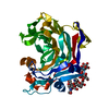
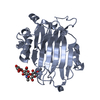
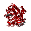
ALFA-LAMINARIHEPTAOSYL FLUORIDE (A401-A407): BETA-1-3-LINKED GLUCAN WITH AN ALPHA-FLUORIDE FOUND IN ...ALFA-LAMINARIHEPTAOSYL FLUORIDE (A401-A407): BETA-1-3-LINKED GLUCAN WITH AN ALPHA-FLUORIDE FOUND IN LAMINARIA DIGITATA. BETA-D-MANNOSE (BMA): PART OF N-GLYCOSYLATION ALPHA-GLYCOSYL-FLUORIDE (GLF): A402 HAS BETA-1,3 GLYCOSIDIC BOND TO THIS RESIDUE IN THE -1 SUBSITE. ALPHA-D-MANNOSE (MAN): PART OF N-GLYCOSYLATION BETA-D-GLUCOSE (BGC): BETA-1,3-GLUCAN IN THE ACTIVE SITE, WITH A407 AT +1 SUBSITE, A406 AT +2 AND A403 AT -3 AND A402 AT -2. B402 IS LINKED TO GLF WITH A BETA-1-3-GLYCOSIDIC BOND. N-ACETYL-D-GLUCOSAMINE (NAG): PART OF N-GLYCOSYLATION
Sequence details
POINT MUTATION OF NUCLEOPHILE RESIDUE GLUTAMATE 115 TO SERINE (THIS IS A E115S MUTANT)
-
Experimental details
-
Experiment
Experiment
Method: X-RAY DIFFRACTION / Number of used crystals: 1
-
Sample preparation
Crystal
Density Matthews: 2 Å3/Da / Density % sol: 40 % / Description: NONE
Crystal grow
Method: vapor diffusion, hanging drop / pH: 5 Details: HANGING DROP: 1 UL 2MG/ML PROTEIN MIXED WITH 1 UL RESERVOIR SOLUTION: 20% PEG 3350, 0.2 M NH4NO3 PH 5 (REERVOIR VOLUME 500 UL). XTALS WERE SOAKED O/N IN DROP OF 10 MM AL7F AND EQUAL VOLUME ...Details: HANGING DROP: 1 UL 2MG/ML PROTEIN MIXED WITH 1 UL RESERVOIR SOLUTION: 20% PEG 3350, 0.2 M NH4NO3 PH 5 (REERVOIR VOLUME 500 UL). XTALS WERE SOAKED O/N IN DROP OF 10 MM AL7F AND EQUAL VOLUME 35% PEG, 0.2 M NH4NO3 PH 5.
-
Data collection
Diffraction
Mean temperature: 100 K
Diffraction source
Source: SYNCHROTRON / Site: MAX II / Beamline: I911-2 / Wavelength: 1.0379
 Movie
Movie Controller


 Open data
 Basic information
 Components
 Keywords
 Function and homology information
 PHANEROCHAETE CHRYSOSPORIUM (fungus)
 X-RAY DIFFRACTION /
  Authors
 Citation
 Structure visualization
 Downloads & links
 Other downloads


 PDBj
PDBj
 Assembly
 Mass: 18.015 Da / Num. of mol.: 263 / Source method: isolated from a natural source / Formula: H2O
 Sample preparation
 / Beamline: I911-2 / Wavelength: 1.0379
  Processing