 Movie
Movie Controller

[English] 日本語
 Yorodumi
- PDB-2wc1: Three-dimensional Structure of the Nitrogen Fixation Flavodoxin (... -
+ Open data

- Basic information
| Entry | Database: PDB / ID: 2wc1 | ||||||
|---|---|---|---|---|---|---|---|
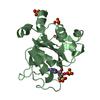
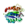
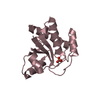
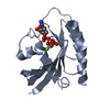
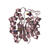
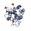
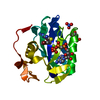
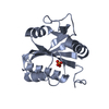
| Title | Three-dimensional Structure of the Nitrogen Fixation Flavodoxin (NifF) from Rhodobacter capsulatus at 2.2 A | ||||||
 Components Components | FLAVODOXIN | ||||||
 Keywords Keywords | ELECTRON TRANSPORT / FLAVOPROTEIN | ||||||
| Function / homology |  Function and homology information Function and homology information | ||||||
| Biological species |  RHODOBACTER CAPSULATUS (bacteria) RHODOBACTER CAPSULATUS (bacteria) | ||||||
| Method |  X-RAY DIFFRACTION / MOLECULAR REPLACEMENT / Resolution: 2.17 Å X-RAY DIFFRACTION / MOLECULAR REPLACEMENT / Resolution: 2.17 Å | ||||||
 Authors Authors | Perez-Dorado, I. / Bittel, C. / Hermoso, J.A. / Cortez, N. / Carrillo, N. | ||||||
 Citation Citation | Journal: Int.J.Mol.Sci. / Year: 2013 Title: Structural and Phylogenetic Analysis of Rhodobacter Capsulatus Niff: Uncovering General Features of Nitrogen-Fixation (Nif)-Flavodoxins. Authors: Perez-Dorado, I. / Bortolotti, A. / Cortez, N. / Hermoso, J.A. #1: Journal: Acta Crystallogr.,Sect.F / Year: 2008 Title: Crystallization of a Flavodoxin Involved in Nitrogen Fixation in Rhodobacter Capsulatus. Authors: Perez-Dorado, I. / Bortolotti, A. / Cortez, N. / Hermoso, J.A. #2: Journal: Biochemistry / Year: 2005Title: The Ferredoxin-Nadp(H) Reductase from Rhodobacter Capsulatus: Molecular Structure and Catalytic Mechanism. Authors: Nogues, I. / Perez-Dorado, I. / Frago, S. / Bittel, C. / Mayhew, S.G. / Gomez-Moreno, C. / Hermoso, J.A. / Medina, M. / Cortez, N. / Carrillo, N. #3: Journal: FEBS Lett. / Year: 2003 Title: The Oxidant-Responsive Diaphorase of Rhodobacter Capsulatus is a Ferredoxin (Flavodoxin)-Nadp(H) Reductase. Authors: Bittel, C. / Tabares, L.C. / Armesto, M. / Carrillo, N. / Cortez, N. #4: Journal: Biochim.Biophys.Acta / Year: 1998 Title: Stopped-Flow Kinetic Studies of Low Potential Electron Carriers of the Photosynthetic Bacterium, Rhodobacter Capsulatus: Ferredoxin I and Niff. Authors: Hallenbeck, P.C. / Gennaro, G. #5: Journal: J.Bacteriol. / Year: 1996 Title: Cloning, Characterization, and Regulation of Niff from Rhodobacter Capsulatus. Authors: Gennaro, G. / Hubner, P. / Sandmeier, U. / Yakunin, A.F. / Hallenbeck, P.C. #6: Journal: J.Bacteriol. / Year: 1993 Title: Purification and Properties of a Nif-Specific Flavodoxin from the Photosynthetic Bacterium Rhodobacter Capsulatus. Authors: Yakunin, A.F. / Gennaro, G. / Hallenbeck, P.C. | ||||||
| History |
| ||||||
| Remark 700 | SHEET THE SHEET STRUCTURE OF THIS MOLECULE IS BIFURCATED. IN ORDER TO REPRESENT THIS FEATURE IN ... SHEET THE SHEET STRUCTURE OF THIS MOLECULE IS BIFURCATED. IN ORDER TO REPRESENT THIS FEATURE IN THE SHEET RECORDS BELOW, TWO SHEETS ARE DEFINED. |
- Structure visualization
| Structure viewer | Molecule: MolmilJmol/JSmol |
|---|
- Downloads & links
-Download
| PDBx/mmCIF format | 2wc1.cif.gz | 51.3 KB | Display | PDBx/mmCIF format |
|---|---|---|---|---|
| PDB format | pdb2wc1.ent.gz | 36 KB | Display | PDB format |
| PDBx/mmJSON format | 2wc1.json.gz | Tree view | PDBx/mmJSON format | |
| Others |  Other downloads Other downloads |
-Validation report
| Arichive directory | https://data.pdbj.org/pub/pdb/validation_reports/wc/2wc1ftp://data.pdbj.org/pub/pdb/validation_reports/wc/2wc1 | HTTPS FTP |
|---|
-Related structure data
| Related structure data | 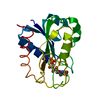 1flvS S: Starting model for refinement |
|---|---|
| Similar structure data |


-Links
 PDBj
PDBj

- Assembly
| Deposited unit | 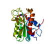
| ||||||||
|---|---|---|---|---|---|---|---|---|---|
| 1 |
| ||||||||
| Unit cell |
|
-Components
| #1: Protein | Mass: 19834.410 Da / Num. of mol.: 1 Source method: isolated from a genetically manipulated source Source: (gene. exp.) RHODOBACTER CAPSULATUS (bacteria) / Plasmid: PET32-FLD / Production host: |
|---|---|
| #2: Chemical | ChemComp-FMN /   Mass: 456.344 Da / Num. of mol.: 1 / Source method: obtained synthetically / Formula: C17H21N4O9P Mass: 456.344 Da / Num. of mol.: 1 / Source method: obtained synthetically / Formula: C17H21N4O9P |
| #3: Water | ChemComp-HOH /  Mass: 18.015 Da / Num. of mol.: 74 / Source method: isolated from a natural source / Formula: H2O Mass: 18.015 Da / Num. of mol.: 74 / Source method: isolated from a natural source / Formula: H2O |
-Experimental details
-Experiment
| Experiment | Method: X-RAY DIFFRACTION / Number of used crystals: 1 |
|---|
- Sample preparation
| Crystal | Density Matthews: 3.4 Å3/Da / Density % sol: 63.7 % / Description: NONE |
|---|
-Data collection
| Diffraction | Mean temperature: 120 K |
|---|---|
| Diffraction source | Source: ROTATING ANODE / Type: ENRAF-NONIUS FR571 / Wavelength: 1.5418 |
| Detector | Type: MARRESEARCH / Detector: IMAGE PLATE / Date: Feb 25, 2004 / Details: MIRRORS |
| Radiation | Monochromator: GRAPHITE / Protocol: SINGLE WAVELENGTH / Monochromatic (M) / Laue (L): M / Scattering type: x-ray |
| Radiation wavelength | Wavelength: 1.5418 Å / Relative weight: 1 |
| Reflection | Resolution: 2.2→25.7 Å / Num. obs: 24830 / % possible obs: 99.9 % / Observed criterion σ(I): 2 / Redundancy: 22.4 % / Biso Wilson estimate: 32.6 Å2 / Rmerge(I) obs: 0.07 / Net I/σ(I): 26 |
| Reflection shell | Resolution: 2.2→2.4 Å / Redundancy: 22.9 % / Rmerge(I) obs: 0.49 / Mean I/σ(I) obs: 8.2 / % possible all: 100 |
- Processing
| Software |
| ||||||||||||||||||||||||||||||||||||||||||||||||||||||||||||||||||||||||||||||||
|---|---|---|---|---|---|---|---|---|---|---|---|---|---|---|---|---|---|---|---|---|---|---|---|---|---|---|---|---|---|---|---|---|---|---|---|---|---|---|---|---|---|---|---|---|---|---|---|---|---|---|---|---|---|---|---|---|---|---|---|---|---|---|---|---|---|---|---|---|---|---|---|---|---|---|---|---|---|---|---|---|---|
| Refinement | Method to determine structure: MOLECULAR REPLACEMENT Starting model: PDB ENTRY 1FLV Resolution: 2.17→25.66 Å / Rfactor Rfree error: 0.008 / Data cutoff high absF: 1425280.34 / Data cutoff low absF: 0 / Isotropic thermal model: RESTRAINED / Cross valid method: THROUGHOUT / σ(F): 0
| ||||||||||||||||||||||||||||||||||||||||||||||||||||||||||||||||||||||||||||||||
| Solvent computation | Solvent model: FLAT MODEL / Bsol: 67.1858 Å2 / ksol: 0.414581 e/Å3 | ||||||||||||||||||||||||||||||||||||||||||||||||||||||||||||||||||||||||||||||||
| Displacement parameters | Biso mean: 43.5 Å2
| ||||||||||||||||||||||||||||||||||||||||||||||||||||||||||||||||||||||||||||||||
| Refine analyze |
| ||||||||||||||||||||||||||||||||||||||||||||||||||||||||||||||||||||||||||||||||
| Refinement step | Cycle: LAST / Resolution: 2.17→25.66 Å
| ||||||||||||||||||||||||||||||||||||||||||||||||||||||||||||||||||||||||||||||||
| Refine LS restraints |
| ||||||||||||||||||||||||||||||||||||||||||||||||||||||||||||||||||||||||||||||||
| Refine LS restraints NCS | NCS model details: NONE | ||||||||||||||||||||||||||||||||||||||||||||||||||||||||||||||||||||||||||||||||
| LS refinement shell | Resolution: 2.17→2.31 Å / Rfactor Rfree error: 0.028 / Total num. of bins used: 6
| ||||||||||||||||||||||||||||||||||||||||||||||||||||||||||||||||||||||||||||||||
| Xplor file |
|
