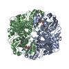
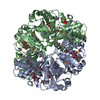
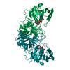
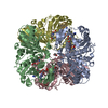
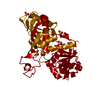
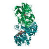
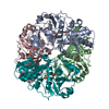
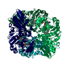
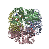
[English] 日本語
Yorodumi
- PDB-2vyn: Structure of E.Coli GAPDH Rat Sperm GAPDH heterotetramer -

+
Open data


ID or keywords:

Loading...

-
Basic information

Entry
Database: PDB / ID: 2vyn
TitleStructure of E.Coli GAPDH Rat Sperm GAPDH heterotetramer
Components(GLYCERALDEHYDE-3-PHOSPHATE DEHYDROGENASEGlyceraldehyde 3-phosphate dehydrogenase) x 2
KeywordsOXIDOREDUCTASE / GAPDH / CONTRACEPTIVE / RAT SPERM / GLYCERALDEHYDE-3-PHOSPHATE / ALPHA CHLOROHYDRIN
Function / homology
Function and homology information


Glycolysis / sperm fibrous sheath / sperm principal piece / Gluconeogenesis / flagellated sperm motility / glyceraldehyde-3-phosphate dehydrogenase (phosphorylating) / glyceraldehyde-3-phosphate dehydrogenase (NAD+) (phosphorylating) activity / motile cilium / spermatid development / acrosomal vesicle ...Glycolysis / sperm fibrous sheath / sperm principal piece / Gluconeogenesis / flagellated sperm motility / glyceraldehyde-3-phosphate dehydrogenase (phosphorylating) / glyceraldehyde-3-phosphate dehydrogenase (NAD+) (phosphorylating) activity / motile cilium / spermatid development / acrosomal vesicle / glycolytic process / cilium / glucose metabolic process / NAD binding / NADP binding / membrane / identical protein binding / cytosol
Similarity search - Function
Glyceraldehyde-3-phosphate dehydrogenase, type I / Glyceraldehyde 3-phosphate dehydrogenase, active site / Glyceraldehyde 3-phosphate dehydrogenase active site. / Glyceraldehyde 3-phosphate dehydrogenase, NAD binding domain / Glyceraldehyde 3-phosphate dehydrogenase, NAD(P) binding domain / Glyceraldehyde 3-phosphate dehydrogenase, catalytic domain / Glyceraldehyde/Erythrose phosphate dehydrogenase family / Glyceraldehyde 3-phosphate dehydrogenase, C-terminal domain / Glyceraldehyde 3-phosphate dehydrogenase, NAD binding domain / Dihydrodipicolinate Reductase; domain 2 ...Glyceraldehyde-3-phosphate dehydrogenase, type I / Glyceraldehyde 3-phosphate dehydrogenase, active site / Glyceraldehyde 3-phosphate dehydrogenase active site. / Glyceraldehyde 3-phosphate dehydrogenase, NAD binding domain / Glyceraldehyde 3-phosphate dehydrogenase, NAD(P) binding domain / Glyceraldehyde 3-phosphate dehydrogenase, catalytic domain / Glyceraldehyde/Erythrose phosphate dehydrogenase family / Glyceraldehyde 3-phosphate dehydrogenase, C-terminal domain / Glyceraldehyde 3-phosphate dehydrogenase, NAD binding domain / Dihydrodipicolinate Reductase; domain 2 / Dihydrodipicolinate Reductase; domain 2 / NAD(P)-binding Rossmann-like Domain / NAD(P)-binding domain superfamily / Rossmann fold / 2-Layer Sandwich / 3-Layer(aba) Sandwich / Alpha Beta
Similarity search - Domain/homology
FORMIC ACID / NICOTINAMIDE-ADENINE-DINUCLEOTIDE / Glyceraldehyde-3-phosphate dehydrogenase A / Glyceraldehyde-3-phosphate dehydrogenase, testis-specific
Similarity search - Component
Biological speciesESCHERICHIA COLI BL21 (bacteria)
RATTUS NORVEGICUS (Norway rat)
MethodX-RAY DIFFRACTION / SYNCHROTRON / MOLECULAR REPLACEMENT / Resolution: 2.2 Å
AuthorsFrayne, J. / Taylor, A. / Hall, L. / Hadfield, A.
CitationJournal: J.Biol.Chem. / Year: 2009
Title: Structure of Insoluble Rat Sperm Glyceraldehyde-3-Phosphate Dehydrogenase (Gapdh) Via Heterotetramer Formation with Escherichia Coli Gapdh Reveals Target for Contraceptive Design.
Authors: Frayne, J. / Taylor, A. / Cameron, G. / Hadfield, A.T.
History
DepositionJul 25, 2008Deposition site: PDBE / Processing site: PDBE
Revision 1.0Aug 5, 2008Provider: repository / Type: Initial release
Revision 1.1May 7, 2011Group: Version format compliance
Revision 1.2Jul 13, 2011Group: Version format compliance
Revision 1.3Oct 31, 2018Group: Data collection / Source and taxonomy / Category: entity_src_nat
Item: _entity_src_nat.pdbx_ncbi_taxonomy_id / _entity_src_nat.pdbx_organism_scientific ..._entity_src_nat.pdbx_ncbi_taxonomy_id / _entity_src_nat.pdbx_organism_scientific / _entity_src_nat.pdbx_variant / _entity_src_nat.strain
Revision 1.4Dec 13, 2023Group: Data collection / Database references ...Data collection / Database references / Derived calculations / Other / Refinement description
Category: chem_comp_atom / chem_comp_bond ...chem_comp_atom / chem_comp_bond / database_2 / pdbx_database_status / pdbx_initial_refinement_model / struct_conn
Item: _database_2.pdbx_DOI / _database_2.pdbx_database_accession ..._database_2.pdbx_DOI / _database_2.pdbx_database_accession / _pdbx_database_status.status_code_sf / _struct_conn.pdbx_leaving_atom_flag
Remark 700 SHEET THE SHEET STRUCTURE OF THIS MOLECULE IS BIFURCATED. IN ORDER TO REPRESENT THIS FEATURE IN ... SHEET THE SHEET STRUCTURE OF THIS MOLECULE IS BIFURCATED. IN ORDER TO REPRESENT THIS FEATURE IN THE SHEET RECORDS BELOW, TWO SHEETS ARE DEFINED.

-
Structure visualization

Structure viewerMolecule:
MolmilJmol/JSmol

Downloads & links

-
Assembly

Deposited unit
A: GLYCERALDEHYDE-3-PHOSPHATE DEHYDROGENASE
B: GLYCERALDEHYDE-3-PHOSPHATE DEHYDROGENASE
C: GLYCERALDEHYDE-3-PHOSPHATE DEHYDROGENASE
D: GLYCERALDEHYDE-3-PHOSPHATE DEHYDROGENASE
hetero molecules


Theoretical massNumber of molelcules
Total (without water)146,59922
Polymers143,3014
Non-polymers3,29818
Water15,205844
1


  • Idetical with deposited unit
  • defined by author&software
TypeNameSymmetry operationNumber
identity operation1_555x,y,z1
Buried area17620 Å2
ΔGint-133 kcal/mol
Surface area54550 Å2
MethodPQS
Unit cell
Length a, b, c (Å)68.445, 103.562, 177.832
Angle α, β, γ (deg.)90.00, 90.00, 90.00
Int Tables number19
Space group name H-MP212121
Noncrystallographic symmetry (NCS)NCS oper:
IDCodeMatrixVector
1given(0.894, 0.435, 0.112), (0.434, -0.9, 0.027), (0.113, 0.025, -0.993)-22.2399, 86.23087, 40.96305
2given(-0.9964, 0.01771, -0.08294), (-0.0032, -0.98511, -0.17187), (-0.08475, -0.17099, 0.98162)46.85013, 105.05235, 11.08776

-
Components

#1: Protein GLYCERALDEHYDE-3-PHOSPHATE DEHYDROGENASE / Glyceraldehyde 3-phosphate dehydrogenase


Mass: 35641.434 Da / Num. of mol.: 3 / Source method: isolated from a natural source / Source: (natural) ESCHERICHIA COLI BL21(DE3) (bacteria) / Variant: pLysS
References: UniProt: P0A9B2, glyceraldehyde-3-phosphate dehydrogenase (phosphorylating)
#2: Protein GLYCERALDEHYDE-3-PHOSPHATE DEHYDROGENASE / Glyceraldehyde 3-phosphate dehydrogenase


Mass: 36376.578 Da / Num. of mol.: 1 / Fragment: RESIDUES 102-432 / Source method: isolated from a natural source / Details: DNA WAS EXTRACTED DIRECTLY FROM RAT TESTIS / Source: (natural) RATTUS NORVEGICUS (Norway rat) / Tissue: TESTISTesticle
References: UniProt: Q9ESV6, glyceraldehyde-3-phosphate dehydrogenase (phosphorylating)
#3: Chemical
ChemComp-FMT / FORMIC ACID / Formic acid


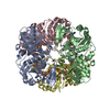
Mass: 46.025 Da / Num. of mol.: 14 / Source method: obtained synthetically / Formula: CH2O2
#4: Chemical
ChemComp-NAD / NICOTINAMIDE-ADENINE-DINUCLEOTIDE / Nicotinamide adenine dinucleotide


Mass: 663.425 Da / Num. of mol.: 4 / Source method: obtained synthetically / Formula: C21H27N7O14P2 / Comment: NAD*YM
#5: Water ChemComp-HOH / water / Water


Mass: 18.015 Da / Num. of mol.: 844 / Source method: isolated from a natural source / Formula: H2O

-
Experimental details

-
Experiment

ExperimentMethod: X-RAY DIFFRACTION / Number of used crystals: 1

-
Sample preparation

CrystalDensity Matthews: 2.1 Å3/Da / Density % sol: 42 % / Description: NONE
Crystal growpH: 7.5 / Details: 0.2M NA FORMATE, 20% PEG 3350, 0.1M HEPES PH 7.5

-
Data collection

DiffractionMean temperature: 100 K
Diffraction sourceSource: SYNCHROTRON / Site: ESRF / Beamline: BM14 / Wavelength: 1
DetectorType: MARRESEARCH / Detector: CCD / Date: Jul 21, 2006
RadiationProtocol: SINGLE WAVELENGTH / Monochromatic (M) / Laue (L): M / Scattering type: x-ray
Radiation wavelengthWavelength: 1 Å / Relative weight: 1
ReflectionResolution: 2.2→25 Å / Num. obs: 63912 / % possible obs: 97.7 % / Observed criterion σ(I): 0 / Redundancy: 4.8 % / Rmerge(I) obs: 0.07 / Net I/σ(I): 21
Reflection shellResolution: 2.2→2.26 Å / Redundancy: 4.7 % / Rmerge(I) obs: 0.23 / Mean I/σ(I) obs: 5 / % possible all: 98.4

-
Processing

Software
NameVersionClassification
REFMAC5.2.0019refinement
HKL-2000data reduction
HKL-2000data scaling
PHASERphasing
RefinementMethod to determine structure: MOLECULAR REPLACEMENT
Starting model: PDB ENTRY 1GAD
Resolution: 2.2→30 Å / Cor.coef. Fo:Fc: 0.958 / Cor.coef. Fo:Fc free: 0.923 / SU B: 5.298 / SU ML: 0.138 / Cross valid method: THROUGHOUT / ESU R: 0.301 / ESU R Free: 0.218 / Stereochemistry target values: MAXIMUM LIKELIHOOD / Details: HYDROGENS HAVE BEEN ADDED IN THE RIDING POSITIONS.
RfactorNum. reflection% reflectionSelection details
Rfree0.224 3224 5.1 %RANDOM
Rwork0.157 ---
obs0.16 60321 97.7 %-
Solvent computationIon probe radii: 0.8 Å / Shrinkage radii: 0.8 Å / VDW probe radii: 1.4 Å / Solvent model: BABINET MODEL WITH MASK
Displacement parametersBiso mean: 25.05 Å2
Baniso -1Baniso -2Baniso -3
1-0.38 Å20 Å20 Å2
2---0.8 Å20 Å2
3---0.42 Å2
Refinement stepCycle: LAST / Resolution: 2.2→30 Å
ProteinNucleic acidLigandSolventTotal
Num. atoms10026 0 218 844 11088
Refine LS restraints
Refine-IDTypeDev idealDev ideal targetNumber
X-RAY DIFFRACTIONr_bond_refined_d0.0090.02210555
X-RAY DIFFRACTIONr_bond_other_d
X-RAY DIFFRACTIONr_angle_refined_deg1.2581.97914337
X-RAY DIFFRACTIONr_angle_other_deg
X-RAY DIFFRACTIONr_dihedral_angle_1_deg6.18251353
X-RAY DIFFRACTIONr_dihedral_angle_2_deg39.57824.753425
X-RAY DIFFRACTIONr_dihedral_angle_3_deg14.527151766
X-RAY DIFFRACTIONr_dihedral_angle_4_deg20.9411552
X-RAY DIFFRACTIONr_chiral_restr0.0840.21659
X-RAY DIFFRACTIONr_gen_planes_refined0.0040.027821
X-RAY DIFFRACTIONr_gen_planes_other
X-RAY DIFFRACTIONr_nbd_refined0.2010.25444
X-RAY DIFFRACTIONr_nbd_other
X-RAY DIFFRACTIONr_nbtor_refined0.3030.27214
X-RAY DIFFRACTIONr_nbtor_other
X-RAY DIFFRACTIONr_xyhbond_nbd_refined0.1520.21061
X-RAY DIFFRACTIONr_xyhbond_nbd_other
X-RAY DIFFRACTIONr_metal_ion_refined
X-RAY DIFFRACTIONr_metal_ion_other
X-RAY DIFFRACTIONr_symmetry_vdw_refined0.1730.242
X-RAY DIFFRACTIONr_symmetry_vdw_other
X-RAY DIFFRACTIONr_symmetry_hbond_refined0.1830.219
X-RAY DIFFRACTIONr_symmetry_hbond_other
X-RAY DIFFRACTIONr_symmetry_metal_ion_refined
X-RAY DIFFRACTIONr_symmetry_metal_ion_other
X-RAY DIFFRACTIONr_mcbond_it1.38936831
X-RAY DIFFRACTIONr_mcbond_other
X-RAY DIFFRACTIONr_mcangle_it2.21510695
X-RAY DIFFRACTIONr_mcangle_other
X-RAY DIFFRACTIONr_scbond_it3.41964204
X-RAY DIFFRACTIONr_scbond_other
X-RAY DIFFRACTIONr_scangle_it4.989103634
X-RAY DIFFRACTIONr_scangle_other
X-RAY DIFFRACTIONr_long_range_B_refined
X-RAY DIFFRACTIONr_long_range_B_other
X-RAY DIFFRACTIONr_rigid_bond_restr
X-RAY DIFFRACTIONr_sphericity_free
X-RAY DIFFRACTIONr_sphericity_bonded
LS refinement shellResolution: 2.2→2.26 Å / Total num. of bins used: 20 /
RfactorNum. reflection
Rfree0.301 244
Rwork0.164 4412

+
About Yorodumi

-
News

-
Feb 9, 2022. New format data for meta-information of EMDB entries

New format data for meta-information of EMDB entries

  • Version 3 of the EMDB header file is now the official format.
  • The previous official version 1.9 will be removed from the archive.

Related info.:EMDB header

External links:wwPDB to switch to version 3 of the EMDB data model

-
Aug 12, 2020. Covid-19 info

Covid-19 info

URL: https://pdbj.org/emnavi/covid19.php

New page: Covid-19 featured information page in EM Navigator.

Related info.:Covid-19 info / Mar 5, 2020. Novel coronavirus structure data

+
Mar 5, 2020. Novel coronavirus structure data

Novel coronavirus structure data

Related info.:Yorodumi Speices / Aug 12, 2020. Covid-19 info

External links:COVID-19 featured content - PDBj / Molecule of the Month (242):Coronavirus Proteases

+
Jan 31, 2019. EMDB accession codes are about to change! (news from PDBe EMDB page)

EMDB accession codes are about to change! (news from PDBe EMDB page)

  • The allocation of 4 digits for EMDB accession codes will soon come to an end. Whilst these codes will remain in use, new EMDB accession codes will include an additional digit and will expand incrementally as the available range of codes is exhausted. The current 4-digit format prefixed with “EMD-” (i.e. EMD-XXXX) will advance to a 5-digit format (i.e. EMD-XXXXX), and so on. It is currently estimated that the 4-digit codes will be depleted around Spring 2019, at which point the 5-digit format will come into force.
  • The EM Navigator/Yorodumi systems omit the EMD- prefix.

Related info.:Q: What is EMD? / ID/Accession-code notation in Yorodumi/EM Navigator

External links:EMDB Accession Codes are Changing Soon! / Contact to PDBj

+
Jul 12, 2017. Major update of PDB

Major update of PDB

  • wwPDB released updated PDB data conforming to the new PDBx/mmCIF dictionary.
  • This is a major update changing the version number from 4 to 5, and with Remediation, in which all the entries are updated.
  • In this update, many items about electron microscopy experimental information are reorganized (e.g. em_software).
  • Now, EM Navigator and Yorodumi are based on the updated data.

External links:wwPDB Remediation / Enriched Model Files Conforming to OneDep Data Standards Now Available in the PDB FTP Archive

-
Yorodumi

Thousand views of thousand structures

  • Yorodumi is a browser for structure data from EMDB, PDB, SASBDB, etc.
  • This page is also the successor to EM Navigator detail page, and also detail information page/front-end page for Omokage search.
  • The word "yorodu" (or yorozu) is an old Japanese word meaning "ten thousand". "mi" (miru) is to see.

Related info.:EMDB / PDB / SASBDB / Comparison of 3 databanks / Yorodumi Search / Aug 31, 2016. New EM Navigator & Yorodumi / Yorodumi Papers / Jmol/JSmol / Function and homology information / Changes in new EM Navigator and Yorodumi

Read more