| Entry | Database: PDB / ID: 2vvd
|
|---|
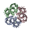
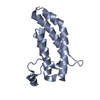
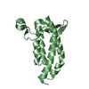
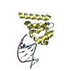
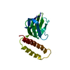
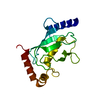
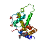
| Title | Crystal structure of the receptor binding domain of the spike protein P1 from bacteriophage PM2 |
|---|
 Components Components | SPIKE PROTEIN P1 |
|---|
 Keywords Keywords | VIRAL PROTEIN / VIRAL RECEPTOR BINDING DOMAIN |
|---|
| Function / homology | Jelly Rolls - #720 / Phage PM2 spike protein P1 / Spike protein P1, first jelly-roll domain / Spike protein P1, second jelly-roll domain / virion component / Jelly Rolls / Sandwich / Mainly Beta / Spike protein P1 Function and homology information Function and homology information |
|---|
| Biological species |  PSEUDOALTEROMONAS PHAGE PM2 (virus) PSEUDOALTEROMONAS PHAGE PM2 (virus) |
|---|
| Method |  X-RAY DIFFRACTION / SYNCHROTRON / MAD / Resolution: 2.26 Å X-RAY DIFFRACTION / SYNCHROTRON / MAD / Resolution: 2.26 Å |
|---|
 Authors Authors | Abrescia, N.G.A. / Grimes, J.M. / Kivela, H.K. / Assenberg, R. / Sutton, G.C. / Butcher, S.J. / Bamford, J.K.H. / Bamford, D.H. / Stuart, D.I. |
|---|
 Citation Citation | Journal: Mol.Cell / Year: 2008
Title: Insights Into Virus Evolution and Membrane Biogenesis from the Structure of the Marine Lipid-Containing Bacteriophage Pm2.
Authors: Abrescia, N.G.A. / Grimes, J.M. / Kivela, H.K. / Assenberg, R. / Sutton, G.C. / Butcher, S.J. / Bamford, J.K.H. / Bamford, D.H. / Stuart, D.I. |
|---|
| History | | Deposition | Jun 6, 2008 | Deposition site: PDBE / Processing site: PDBE |
|---|
| Revision 1.0 | Sep 16, 2008 | Provider: repository / Type: Initial release |
|---|
| Revision 1.1 | May 8, 2011 | Group: Version format compliance |
|---|
| Revision 1.2 | Jul 13, 2011 | Group: Version format compliance |
|---|
| Revision 1.3 | Oct 23, 2024 | Group: Data collection / Database references ...Data collection / Database references / Derived calculations / Other / Structure summary
Category: chem_comp_atom / chem_comp_bond ...chem_comp_atom / chem_comp_bond / database_2 / pdbx_database_status / pdbx_entry_details / pdbx_modification_feature / pdbx_struct_conn_angle / struct_conn
Item: _database_2.pdbx_DOI / _database_2.pdbx_database_accession ..._database_2.pdbx_DOI / _database_2.pdbx_database_accession / _pdbx_database_status.status_code_sf / _pdbx_entry_details.has_protein_modification / _pdbx_struct_conn_angle.ptnr1_auth_comp_id / _pdbx_struct_conn_angle.ptnr1_auth_seq_id / _pdbx_struct_conn_angle.ptnr1_label_asym_id / _pdbx_struct_conn_angle.ptnr1_label_atom_id / _pdbx_struct_conn_angle.ptnr1_label_comp_id / _pdbx_struct_conn_angle.ptnr1_label_seq_id / _pdbx_struct_conn_angle.ptnr1_symmetry / _pdbx_struct_conn_angle.ptnr2_auth_seq_id / _pdbx_struct_conn_angle.ptnr2_label_asym_id / _pdbx_struct_conn_angle.ptnr3_auth_comp_id / _pdbx_struct_conn_angle.ptnr3_auth_seq_id / _pdbx_struct_conn_angle.ptnr3_label_asym_id / _pdbx_struct_conn_angle.ptnr3_label_atom_id / _pdbx_struct_conn_angle.ptnr3_label_comp_id / _pdbx_struct_conn_angle.ptnr3_label_seq_id / _pdbx_struct_conn_angle.ptnr3_symmetry / _pdbx_struct_conn_angle.value / _struct_conn.pdbx_dist_value / _struct_conn.pdbx_leaving_atom_flag / _struct_conn.ptnr1_auth_comp_id / _struct_conn.ptnr1_auth_seq_id / _struct_conn.ptnr1_label_asym_id / _struct_conn.ptnr1_label_atom_id / _struct_conn.ptnr1_label_comp_id / _struct_conn.ptnr1_label_seq_id / _struct_conn.ptnr1_symmetry / _struct_conn.ptnr2_auth_comp_id / _struct_conn.ptnr2_auth_seq_id / _struct_conn.ptnr2_label_asym_id / _struct_conn.ptnr2_label_atom_id / _struct_conn.ptnr2_label_comp_id / _struct_conn.ptnr2_label_seq_id / _struct_conn.ptnr2_symmetry |
|---|
|
|---|
 Movie
Movie Controller

 Yorodumi
 Open data
 Basic information
 Structure visualization
 Downloads & links
 Other downloads
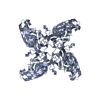


 PDBj
PDBj Assembly
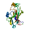


 Mass: 40.078 Da / Num. of mol.: 2 / Source method: obtained synthetically / Formula: Ca
 Mass: 18.015 Da / Num. of mol.: 169 / Source method: isolated from a natural source / Formula: H2O
 Sample preparation
 / Beamline: BM14 / Wavelength: 0.97912,0.90499
 Processing