 Movie
Movie Controller

[English] 日本語
 Yorodumi
- PDB-2vve: Crystal structure of the stem and receptor binding domain of the ... -
+ Open data

- Basic information
| Entry | Database: PDB / ID: 2vve | ||||||
|---|---|---|---|---|---|---|---|
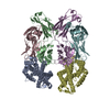
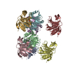
| Title | Crystal structure of the stem and receptor binding domain of the spike protein P1 from bacteriophage PM2 | ||||||
 Components Components | SPIKE PROTEIN P1 | ||||||
 Keywords Keywords | VIRAL PROTEIN / VIRAL STEM-RECEPTOR BINDING DOMAIN | ||||||
| Function / homology |  Function and homology information Function and homology information | ||||||
| Biological species |  PSEUDOALTEROMONAS PHAGE PM2 (virus) PSEUDOALTEROMONAS PHAGE PM2 (virus) | ||||||
| Method |  X-RAY DIFFRACTION / SYNCHROTRON / MOLECULAR REPLACEMENT / Resolution: 1.77 Å X-RAY DIFFRACTION / SYNCHROTRON / MOLECULAR REPLACEMENT / Resolution: 1.77 Å | ||||||
 Authors Authors | Abrescia, N.G.A. / Grimes, J.M. / Kivela, H.K. / Assenberg, R. / Sutton, G.C. / Butcher, S.J. / Bamford, J.K.H. / Bamford, D.H. / Stuart, D.I. | ||||||
 Citation Citation | Journal: Mol.Cell / Year: 2008 Title: Insights Into Virus Evolution and Membrane Biogenesis from the Structure of the Marine Lipid-Containing Bacteriophage Pm2. Authors: Abrescia, N.G.A. / Grimes, J.M. / Kivela, H.K. / Assenberg, R. / Sutton, G.C. / Butcher, S.J. / Bamford, J.K.H. / Bamford, D.H. / Stuart, D.I. | ||||||
| History |
|
- Structure visualization
| Structure viewer | Molecule: MolmilJmol/JSmol |
|---|
- Downloads & links
-Download
| PDBx/mmCIF format | 2vve.cif.gz | 223.3 KB | Display | PDBx/mmCIF format |
|---|---|---|---|---|
| PDB format | pdb2vve.ent.gz | 179 KB | Display | PDB format |
| PDBx/mmJSON format | 2vve.json.gz | Tree view | PDBx/mmJSON format | |
| Others |  Other downloads Other downloads |
-Validation report
| Arichive directory | https://data.pdbj.org/pub/pdb/validation_reports/vv/2vveftp://data.pdbj.org/pub/pdb/validation_reports/vv/2vve | HTTPS FTP |
|---|
-Related structure data
| Related structure data | 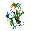 2vvdSC 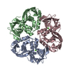 2vvfC 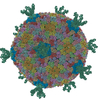 2w0cC S: Starting model for refinement C: citing same article ( |
|---|---|
| Similar structure data |


-Links
 PDBj
PDBj- Assembly
| Deposited unit | 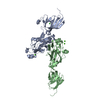
| ||||||||||||
|---|---|---|---|---|---|---|---|---|---|---|---|---|---|
| 1 | 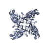
| ||||||||||||
| 2 | 
| ||||||||||||
| Unit cell |
| ||||||||||||
| Components on special symmetry positions |
|
-Components
| #1: Protein | Mass: 28591.408 Da / Num. of mol.: 2 / Fragment: STEM-RECEPTOR BINDING DOMAIN, RESIDUES 82-335 Source method: isolated from a genetically manipulated source Details: CLONED FRAGMENT STARTS AT RESIDUE 82 AND STOPS AT RESIDUE 335 Source: (gene. exp.) PSEUDOALTEROMONAS PHAGE PM2 (virus) / Plasmid: POPINF / Production host:  #2: Chemical | ChemComp-CA /   Mass: 40.078 Da / Num. of mol.: 4 / Source method: obtained synthetically / Formula: Ca Mass: 40.078 Da / Num. of mol.: 4 / Source method: obtained synthetically / Formula: Ca#3: Chemical | ChemComp-CL / |   Mass: 35.453 Da / Num. of mol.: 1 / Source method: obtained synthetically / Formula: Cl Mass: 35.453 Da / Num. of mol.: 1 / Source method: obtained synthetically / Formula: Cl#4: Water | ChemComp-HOH / |  Mass: 18.015 Da / Num. of mol.: 539 / Source method: isolated from a natural source / Formula: H2O Mass: 18.015 Da / Num. of mol.: 539 / Source method: isolated from a natural source / Formula: H2O |
|---|
-Experimental details
-Experiment
| Experiment | Method: X-RAY DIFFRACTION / Number of used crystals: 1 |
|---|
- Sample preparation
| Crystal | Density Matthews: 2.24 Å3/Da / Density % sol: 45.4 % Description: THE ELECTRON DENSITY MAP FOR THE STEM DOMAIN WAS GREATLY IMPROVED BY USING BUSTER PROGRAM IN THE EARLY REFINEMENT CYCLES |
|---|---|
| Crystal grow | pH: 8 Details: 7.3MG/ML (IN 150MM NACL, 20MM TRIS PH 7.5, 10MM CACL2),20%(W/V) PEG6K,100MM TRIS PH 8, 200MM LICL |
-Data collection
| Diffraction | Mean temperature: 100 K |
|---|---|
| Diffraction source | Source: SYNCHROTRON / Site: ESRF  / Beamline: BM14 / Wavelength: 0.9185 / Beamline: BM14 / Wavelength: 0.9185 |
| Detector | Type: MARRESEARCH / Detector: CCD / Date: Aug 30, 2006 / Details: MIRRORS |
| Radiation | Protocol: SINGLE WAVELENGTH / Monochromatic (M) / Laue (L): M / Scattering type: x-ray |
| Radiation wavelength | Wavelength: 0.9185 Å / Relative weight: 1 |
| Reflection | Resolution: 1.77→23 Å / Num. obs: 47548 / % possible obs: 94.9 % / Observed criterion σ(I): -3 / Redundancy: 11.2 % / Biso Wilson estimate: 21.1 Å2 / Rmerge(I) obs: 0.08 / Net I/σ(I): 30.2 |
| Reflection shell | Resolution: 1.77→1.83 Å / Redundancy: 7.1 % / Rmerge(I) obs: 0.87 / Mean I/σ(I) obs: 1.8 / % possible all: 71.8 |
- Processing
| Software |
| ||||||||||||||||||||||||||||||||||||||||||||||||||||||||||||||||||||||||||||||||||||||||||||||||||||||||||||||||||||||||||||||||||||||||||||||||||||||||||||||||||||||||||||||||||||||
|---|---|---|---|---|---|---|---|---|---|---|---|---|---|---|---|---|---|---|---|---|---|---|---|---|---|---|---|---|---|---|---|---|---|---|---|---|---|---|---|---|---|---|---|---|---|---|---|---|---|---|---|---|---|---|---|---|---|---|---|---|---|---|---|---|---|---|---|---|---|---|---|---|---|---|---|---|---|---|---|---|---|---|---|---|---|---|---|---|---|---|---|---|---|---|---|---|---|---|---|---|---|---|---|---|---|---|---|---|---|---|---|---|---|---|---|---|---|---|---|---|---|---|---|---|---|---|---|---|---|---|---|---|---|---|---|---|---|---|---|---|---|---|---|---|---|---|---|---|---|---|---|---|---|---|---|---|---|---|---|---|---|---|---|---|---|---|---|---|---|---|---|---|---|---|---|---|---|---|---|---|---|---|---|
| Refinement | Method to determine structure: MOLECULAR REPLACEMENT Starting model: PDB ENTRY 2VVD Resolution: 1.77→23 Å / Cor.coef. Fo:Fc: 0.96 / Cor.coef. Fo:Fc free: 0.934 / SU B: 5.082 / SU ML: 0.087 / Cross valid method: THROUGHOUT / ESU R: 0.13 / ESU R Free: 0.131 / Stereochemistry target values: MAXIMUM LIKELIHOOD Details: HYDROGENS HAVE BEEN ADDED IN THE RIDING POSITIONS. DESPITE HAVING TWO MOLECULES IN THE ASU THE RESPECTIVE STEM DOMAINS FLEX WITH DIFFERENT ANGLES RELATIVE TO THE RECEPTOR DOMAIN. THEREFORE NO STRICT NCS
| ||||||||||||||||||||||||||||||||||||||||||||||||||||||||||||||||||||||||||||||||||||||||||||||||||||||||||||||||||||||||||||||||||||||||||||||||||||||||||||||||||||||||||||||||||||||
| Solvent computation | Ion probe radii: 0.8 Å / Shrinkage radii: 0.8 Å / VDW probe radii: 1.4 Å / Solvent model: MASK | ||||||||||||||||||||||||||||||||||||||||||||||||||||||||||||||||||||||||||||||||||||||||||||||||||||||||||||||||||||||||||||||||||||||||||||||||||||||||||||||||||||||||||||||||||||||
| Displacement parameters | Biso mean: 18.65 Å2
| ||||||||||||||||||||||||||||||||||||||||||||||||||||||||||||||||||||||||||||||||||||||||||||||||||||||||||||||||||||||||||||||||||||||||||||||||||||||||||||||||||||||||||||||||||||||
| Refinement step | Cycle: LAST / Resolution: 1.77→23 Å
| ||||||||||||||||||||||||||||||||||||||||||||||||||||||||||||||||||||||||||||||||||||||||||||||||||||||||||||||||||||||||||||||||||||||||||||||||||||||||||||||||||||||||||||||||||||||
| Refine LS restraints |
|