- PDB-2vtf: X-ray crystal structure of the Endo-beta-N-acetylglucosaminidase ... -
+
Open data
ID or keywords:
Loading...
-
Basic information
Entry
Database: PDB / ID: 2vtf
Title
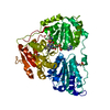
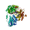
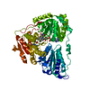
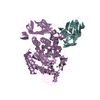
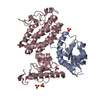
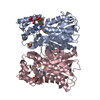
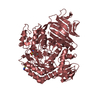
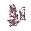
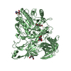
X-ray crystal structure of the Endo-beta-N-acetylglucosaminidase from Arthrobacter protophormiae E173Q mutant reveals a TIM barrel catalytic domain and two ancillary domains
mannosyl-glycoprotein endo-beta-N-acetylglucosaminidase / mannosyl-glycoprotein endo-beta-N-acetylglucosaminidase activity / carbohydrate metabolic process / metal ion binding / cytoplasm Similarity search - Function
Journal: J.Mol.Biol. / Year: 2009 Title: The X-Ray Crystal Structure of an Arthrobacter Protophormiae Endo-Beta-N-Acetylglucosaminidase Reveals a (Beta/Alpha)(8) Catalytic Domain, Two Ancillary Domains and Active Site Residues Key ...Title: The X-Ray Crystal Structure of an Arthrobacter Protophormiae Endo-Beta-N-Acetylglucosaminidase Reveals a (Beta/Alpha)(8) Catalytic Domain, Two Ancillary Domains and Active Site Residues Key for Transglycosylation Activity. Authors: Suits, M.D. / Ling, Z. / Bingham, R.J. / Bruce, N.C. / Davies, G.J. / Fairbanks, A.J. / Moir, J.W. / Taylor, E.J.
SHEET DETERMINATION METHOD: DSSP THE SHEETS PRESENTED AS "AB" IN EACH CHAIN ON SHEET RECORDS BELOW ... SHEET DETERMINATION METHOD: DSSP THE SHEETS PRESENTED AS "AB" IN EACH CHAIN ON SHEET RECORDS BELOW IS ACTUALLY AN 9-STRANDED BARREL THIS IS REPRESENTED BY A 10-STRANDED SHEET IN WHICH THE FIRST AND LAST STRANDS ARE IDENTICAL. THE SHEETS PRESENTED AS "BB" IN EACH CHAIN ON SHEET RECORDS BELOW IS ACTUALLY AN 9-STRANDED BARREL THIS IS REPRESENTED BY A 10-STRANDED SHEET IN WHICH THE FIRST AND LAST STRANDS ARE IDENTICAL. THE SHEET STRUCTURE OF THIS MOLECULE IS BIFURCATED. IN ORDER TO REPRESENT THIS FEATURE IN THE SHEET RECORDS BELOW, TWO SHEETS ARE DEFINED.
Mass: 18.015 Da / Num. of mol.: 1500 / Source method: isolated from a natural source / Formula: H2O
Compound details
ENGINEERED RESIDUE IN CHAIN A, ASN 67 TO ASP ENGINEERED RESIDUE IN CHAIN A, LYS 75 TO THR ...ENGINEERED RESIDUE IN CHAIN A, ASN 67 TO ASP ENGINEERED RESIDUE IN CHAIN A, LYS 75 TO THR ENGINEERED RESIDUE IN CHAIN A, GLU 197 TO GLN ENGINEERED RESIDUE IN CHAIN B, ASN 67 TO ASP ENGINEERED RESIDUE IN CHAIN B, LYS 75 TO THR ENGINEERED RESIDUE IN CHAIN B, GLU 197 TO GLN
-
Experimental details
-
Experiment
Experiment
Method: X-RAY DIFFRACTION / Number of used crystals: 1
-
Sample preparation
Crystal
Density Matthews: 2.52 Å3/Da / Density % sol: 51.22 % / Description: NONE
Crystal grow
pH: 6.5 Details: 0.35 M KSCN, 0.1 M BIS-TRIS PROPANE (PH 6.5), 15% PEG 3350
Monochromator: SI111 CHANNEL CUT / Protocol: SINGLE WAVELENGTH / Monochromatic (M) / Laue (L): M / Scattering type: x-ray
Radiation wavelength
Wavelength: 0.9765 Å / Relative weight: 1
Reflection
Resolution: 1.8→80 Å / Num. obs: 130527 / % possible obs: 99.9 % / Observed criterion σ(I): 5 / Redundancy: 5.7 % / Rmerge(I) obs: 0.09 / Net I/σ(I): 23
Reflection shell
Resolution: 1.8→1.86 Å / Redundancy: 5.3 % / Rmerge(I) obs: 0.34 / Mean I/σ(I) obs: 2.7 / % possible all: 99.7
-
Processing
Software
Name
Version
Classification
REFMAC
5.4.0077
refinement
HKL-2000
datareduction
SCALEPACK
datascaling
autoSHARP
phasing
SOLOMON
phasing
RESOLVE
phasing
Refinement
Method to determine structure: MAD Starting model: NONE Resolution: 1.79→49.03 Å / Cor.coef. Fo:Fc: 0.963 / Cor.coef. Fo:Fc free: 0.943 / SU B: 4.814 / SU ML: 0.069 / TLS residual ADP flag: LIKELY RESIDUAL / Cross valid method: THROUGHOUT / ESU R: 0.113 / ESU R Free: 0.111 / Stereochemistry target values: MAXIMUM LIKELIHOOD Details: HYDROGENS HAVE BEEN ADDED IN THE RIDING POSITIONS. SER207 IS A RAMACHANDRAN PLOT OUTLIER AND IMPORTANT ACTIVE SITE RESIDUE THAT INTERACTS WITH THE CATALYTIC ACID/BASE.
Rfactor
Num. reflection
% reflection
Selection details
Rfree
0.202
6652
5 %
RANDOM
Rwork
0.164
-
-
-
obs
0.166
125475
99.9 %
-
Solvent computation
Ion probe radii: 0.8 Å / Shrinkage radii: 0.8 Å / VDW probe radii: 1.2 Å / Solvent model: MASK
 Movie
Movie Controller

 Yorodumi
 Open data
 Basic information
 Components
 Keywords
 Function and homology information
 ARTHROBACTER PROTOPHORMIAE (bacteria)
 X-RAY DIFFRACTION /
  Authors
 Citation
 Structure visualization
 Downloads & links
 Other downloads
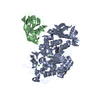

 PDBj
PDBj Assembly
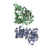


 Mass: 282.334 Da / Num. of mol.: 4 / Source method: obtained synthetically / Formula: C11H26N2O6 / Comment: pH buffer*YM
 Mass: 150.173 Da / Num. of mol.: 4 / Source method: obtained synthetically / Formula: C6H14O4
 Mass: 18.015 Da / Num. of mol.: 1500 / Source method: isolated from a natural source / Formula: H2O
 Sample preparation
 / Beamline: ID14-4 / Wavelength: 0.9765
  Processing