 Movie
Movie Controller

[English] 日本語
 Yorodumi
- PDB-2vt1: Crystal structure of the cytoplasmic domain of Spa40, the specifi... -
+ Open data

- Basic information
| Entry | Database: PDB / ID: 2vt1 | ||||||
|---|---|---|---|---|---|---|---|
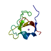
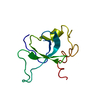
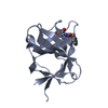
| Title | Crystal structure of the cytoplasmic domain of Spa40, the specificity switch for the Shigella flexneri Type III Secretion System | ||||||
 Components Components | (SURFACE PRESENTATION OF ANTIGENS PROTEIN SPAS) x 2 | ||||||
 Keywords Keywords | MEMBRANE PROTEIN / SHIGELLA FLEXNERI / SPECIFICITY SWITCH / VIRULENCE / TRANSMEMBRANE / INNER MEMBRANE / FLHB / YSCU / T3SS / SPA40 / PLASMID / MEMBRANE / TYPE III SECRETION SYSTEM | ||||||
| Function / homology |  Function and homology information Function and homology information | ||||||
| Biological species |  SHIGELLA FLEXNERI (bacteria) SHIGELLA FLEXNERI (bacteria) | ||||||
| Method |  X-RAY DIFFRACTION / SYNCHROTRON / MOLECULAR REPLACEMENT / Resolution: 2 Å X-RAY DIFFRACTION / SYNCHROTRON / MOLECULAR REPLACEMENT / Resolution: 2 Å | ||||||
 Authors Authors | Deane, J.E. / Graham, S.C. / Mitchell, E.P. / Flot, D. / Johnson, S. / Lea, S.M. | ||||||
 Citation Citation | Journal: Mol.Microbiol. / Year: 2008 Title: Crystal Structure of Spa40, the Specificity Switch for the Shigella Flexneri Type III Secretion System Authors: Deane, J.E. / Graham, S.C. / Mitchell, E.P. / Flot, D. / Johnson, S. / Lea, S.M. | ||||||
| History |
|
- Structure visualization
| Structure viewer | Molecule: MolmilJmol/JSmol |
|---|
- Downloads & links
-Download
| PDBx/mmCIF format | 2vt1.cif.gz | 35 KB | Display | PDBx/mmCIF format |
|---|---|---|---|---|
| PDB format | pdb2vt1.ent.gz | 22.5 KB | Display | PDB format |
| PDBx/mmJSON format | 2vt1.json.gz | Tree view | PDBx/mmJSON format | |
| Others |  Other downloads Other downloads |
-Validation report
| Arichive directory | https://data.pdbj.org/pub/pdb/validation_reports/vt/2vt1ftp://data.pdbj.org/pub/pdb/validation_reports/vt/2vt1 | HTTPS FTP |
|---|
-Related structure data
| Related structure data | 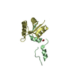 3bzlS S: Starting model for refinement |
|---|---|
| Similar structure data |


-Links
 PDBj
PDBj- Assembly
| Deposited unit | 
| ||||||||
|---|---|---|---|---|---|---|---|---|---|
| 1 |
| ||||||||
| Unit cell |
|
-Components
| #1: Protein | Mass: 6394.362 Da / Num. of mol.: 1 / Fragment: CYTOPLASMIC DOMAIN, RESIDUES 207-257 Source method: isolated from a genetically manipulated source Details: A SINGLE CHAIN OF THE CYTOPLASMIC DOMAIN OF SPA40 IS PRESENT, HAVING UNDERGONE AN AUTO-CLEAVAGE EVENT BETWEEN N257 AND P258 TO FORM TWO CHAINS Source: (gene. exp.) SHIGELLA FLEXNERI (bacteria) / Plasmid: PET-28B / Production host: |
|---|---|
| #2: Protein | Mass: 10890.471 Da / Num. of mol.: 1 / Fragment: CYTOPLASMIC DOMAIN, RESIDUES 258-342 Source method: isolated from a genetically manipulated source Details: A SINGLE CHAIN OF THE CYTOPLASMIC DOMAIN OF SPA40 IS PRESENT, HAVING UNDERGONE AN AUTO-CLEAVAGE EVENT BETWEEN N257 AND P258 TO FORM TWO CHAINS Source: (gene. exp.) SHIGELLA FLEXNERI (bacteria) / Plasmid: PET-28B / Production host: |
| #3: Water | ChemComp-HOH /  Mass: 18.015 Da / Num. of mol.: 29 / Source method: isolated from a natural source / Formula: H2O Mass: 18.015 Da / Num. of mol.: 29 / Source method: isolated from a natural source / Formula: H2O |
| Sequence details | CYTOPLASMI |
-Experimental details
-Experiment
| Experiment | Method: X-RAY DIFFRACTION / Number of used crystals: 1 |
|---|
- Sample preparation
| Crystal | Density Matthews: 1.67 Å3/Da / Density % sol: 26.55 % / Description: NONE |
|---|---|
| Crystal grow | Temperature: 293 K / Method: vapor diffusion, sitting drop / pH: 7 Details: SITTING DROPS CONTAINING 200 NL PROTEIN (3.3 MG/ML IN 20 MM TRIS PH 8.0, 500 MM NACL) AND 200 NL RESERVOIR SOLUTION (0.1M HEPES PH7.0, 0.2M NH4CL AND 20% PEG W/V 6000) WERE EQUILIBRATED ...Details: SITTING DROPS CONTAINING 200 NL PROTEIN (3.3 MG/ML IN 20 MM TRIS PH 8.0, 500 MM NACL) AND 200 NL RESERVOIR SOLUTION (0.1M HEPES PH7.0, 0.2M NH4CL AND 20% PEG W/V 6000) WERE EQUILIBRATED AGAINST 100 UL RESERVOIRS AT 20 C. CRYSTALS WERE CRYOPROTECTED IN RESERVOIR SOLUTION SUPPLEMENTED WITH 25% (V/V) GLYCEROL. THE ASYMMETRIC UNIT VOLUME IS NOT SUFFICIENT TO ACCOMMODATE THE ENTIRE SPA40 CONSTRUCT. WE ASSUME THAT THE PROTEIN HAS UNDERGONE SOME PROTEOLYSIS ADDITIONAL TO THE SELF-CLEAVAGE BETWEEN RESIDUES 257 AND 258, REMOVING EITHER THE DISORDERED N-TERMINUS OR THE C-TERMINAL HIS TAG. THE SOLVENT CONTENT QUOTED IS FOR RESIDUE 237 TO THE END OF THE CONSTRUCT. |
-Data collection
| Diffraction | Mean temperature: 100 K |
|---|---|
| Diffraction source | Source: SYNCHROTRON / Site: ESRF  / Beamline: ID23-1 / Wavelength: 1.0401 / Beamline: ID23-1 / Wavelength: 1.0401 |
| Detector | Type: ADSC CCD / Detector: CCD / Date: Apr 13, 2008 / Details: MIRRORS |
| Radiation | Monochromator: SI(111) CRYSTAL / Protocol: SINGLE WAVELENGTH / Monochromatic (M) / Laue (L): M / Scattering type: x-ray |
| Radiation wavelength | Wavelength: 1.0401 Å / Relative weight: 1 |
| Reflection | Resolution: 2→30 Å / Num. obs: 5433 / % possible obs: 93.2 % / Observed criterion σ(I): -3 / Redundancy: 2.6 % / Biso Wilson estimate: 21.6 Å2 / Rmerge(I) obs: 0.11 / Net I/σ(I): 3.8 |
| Reflection shell | Resolution: 2→2.05 Å / Redundancy: 2.7 % / Rmerge(I) obs: 0.4 / Mean I/σ(I) obs: 1.7 / % possible all: 93.3 |
- Processing
| Software |
| ||||||||||||||||||||||||||||||||||||||||||||||||||||||||||||||||||||||||||||||||||||||||||||||||||||||||||||||||||||||||||||||||||||||||||||||||||||||||||||||||||||||||||||||||||||||
|---|---|---|---|---|---|---|---|---|---|---|---|---|---|---|---|---|---|---|---|---|---|---|---|---|---|---|---|---|---|---|---|---|---|---|---|---|---|---|---|---|---|---|---|---|---|---|---|---|---|---|---|---|---|---|---|---|---|---|---|---|---|---|---|---|---|---|---|---|---|---|---|---|---|---|---|---|---|---|---|---|---|---|---|---|---|---|---|---|---|---|---|---|---|---|---|---|---|---|---|---|---|---|---|---|---|---|---|---|---|---|---|---|---|---|---|---|---|---|---|---|---|---|---|---|---|---|---|---|---|---|---|---|---|---|---|---|---|---|---|---|---|---|---|---|---|---|---|---|---|---|---|---|---|---|---|---|---|---|---|---|---|---|---|---|---|---|---|---|---|---|---|---|---|---|---|---|---|---|---|---|---|---|---|
| Refinement | Method to determine structure: MOLECULAR REPLACEMENT Starting model: PDB ENTRY 3BZL WITH SIDECHAINS MUTATED TO SER Resolution: 2→29.59 Å / Cor.coef. Fo:Fc: 0.958 / Cor.coef. Fo:Fc free: 0.931 / SU B: 5.042 / SU ML: 0.135 / Cross valid method: THROUGHOUT / ESU R: 0.251 / ESU R Free: 0.19 / Stereochemistry target values: MAXIMUM LIKELIHOOD Details: HYDROGENS HAVE BEEN ADDED IN THE RIDING POSITIONS. U VALUES HAVE BEEN REFINED INDIVIDUALLY
| ||||||||||||||||||||||||||||||||||||||||||||||||||||||||||||||||||||||||||||||||||||||||||||||||||||||||||||||||||||||||||||||||||||||||||||||||||||||||||||||||||||||||||||||||||||||
| Solvent computation | Ion probe radii: 0.8 Å / Shrinkage radii: 0.8 Å / VDW probe radii: 1.2 Å / Solvent model: MASK | ||||||||||||||||||||||||||||||||||||||||||||||||||||||||||||||||||||||||||||||||||||||||||||||||||||||||||||||||||||||||||||||||||||||||||||||||||||||||||||||||||||||||||||||||||||||
| Displacement parameters | Biso mean: 18.59 Å2
| ||||||||||||||||||||||||||||||||||||||||||||||||||||||||||||||||||||||||||||||||||||||||||||||||||||||||||||||||||||||||||||||||||||||||||||||||||||||||||||||||||||||||||||||||||||||
| Refinement step | Cycle: LAST / Resolution: 2→29.59 Å
| ||||||||||||||||||||||||||||||||||||||||||||||||||||||||||||||||||||||||||||||||||||||||||||||||||||||||||||||||||||||||||||||||||||||||||||||||||||||||||||||||||||||||||||||||||||||
| Refine LS restraints |
|