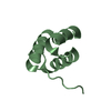
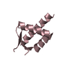
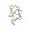
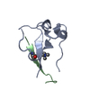
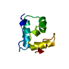
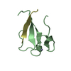
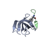
Entry Database : PDB / ID : 2vqcTitle Structure of a DNA binding winged-helix protein, F-112, from Sulfolobus Spindle-shaped Virus 1. HYPOTHETICAL 13.2 KDA PROTEIN Keywords / / / / / / Function / homology / / / / / / / / Biological species Method / / / Resolution : 2.3 Å Authors Menon, S.K. / Kraft, P. / Corn, G.J. / Wiedenheft, B. / Young, M.J. / Lawrence, C.M. Journal : Virology / Year : 2008Title : Cysteine Usage in Sulfolobus Spindle-Shaped Virus 1 and Extension to Hyperthermophilic Viruses in General.Authors : Menon, S.K. / Maaty, W.S. / Corn, G.J. / Kwok, S.C. / Eilers, B.J. / Kraft, P. / Gillitzer, E. / Young, M.J. / Bothner, B. / Lawrence, C.M. History Deposition Mar 12, 2008 Deposition site / Processing site Supersession May 6, 2008 ID 2CMX Revision 1.0 May 6, 2008 Provider / Type Revision 1.1 Jul 13, 2011 Group / Version format complianceRevision 1.2 May 8, 2019 Group Data collection / Derived calculations ... Data collection / Derived calculations / Experimental preparation / Other Category database_PDB_rev / database_PDB_rev_record ... database_PDB_rev / database_PDB_rev_record / exptl_crystal_grow / pdbx_database_proc / pdbx_database_status / struct_conn Item / _pdbx_database_status.recvd_author_approval / _struct_conn.pdbx_leaving_atom_flagRevision 1.3 Nov 20, 2024 Group / Database references / Structure summaryCategory chem_comp_atom / chem_comp_bond ... chem_comp_atom / chem_comp_bond / database_2 / pdbx_entry_details / pdbx_modification_feature Item / _database_2.pdbx_database_accession
Show all Show less
 Movie
Movie Controller

 Yorodumi
 Open data
 Basic information
 Components
 Keywords
 Function and homology information
 SULFOLOBUS VIRUS-LIKE PARTICLE SSV1
 X-RAY DIFFRACTION /
  Authors
 Citation
 Structure visualization
 Downloads & links
 Other downloads
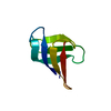


 PDBj
PDBj Assembly
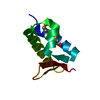

 Mass: 18.015 Da / Num. of mol.: 25 / Source method: isolated from a natural source / Formula: H2O
 Sample preparation
 / Beamline: BL9-2 / Wavelength: 0.891940, 0.979180, 0.979320
 Processing