 Movie
Movie Controller


+ Open data

- Basic information
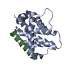
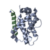
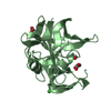
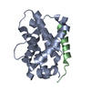
| Entry | Database: PDB / ID: 2vog | ||||||
|---|---|---|---|---|---|---|---|
| Title | Structure of mouse A1 bound to the Bmf BH3-domain | ||||||
 Components Components |
| ||||||
 Keywords Keywords | APOPTOSIS / PROTEIN-PROTEIN COMPLEX / BH3 / BCL-2 / PRO-SURVIVAL | ||||||
| Function / homology |  Function and homology information Function and homology informationActivation of BMF and translocation to mitochondria / BH3-only proteins associate with and inactivate anti-apoptotic BCL-2 members / negative regulation of autophagic cell death / B cell apoptotic process / BH domain binding / negative regulation of B cell apoptotic process / myosin complex / anoikis / channel activity / mitochondrial fusion ...Activation of BMF and translocation to mitochondria / BH3-only proteins associate with and inactivate anti-apoptotic BCL-2 members / negative regulation of autophagic cell death / B cell apoptotic process / BH domain binding / negative regulation of B cell apoptotic process / myosin complex / anoikis / channel activity / mitochondrial fusion / positive regulation of release of cytochrome c from mitochondria / B cell homeostasis / negative regulation of apoptotic signaling pathway / extrinsic apoptotic signaling pathway in absence of ligand / cellular response to starvation / negative regulation of autophagy / release of cytochrome c from mitochondria / acrosomal vesicle / positive regulation of apoptotic signaling pathway / positive regulation of protein-containing complex assembly / cellular response to UV / intrinsic apoptotic signaling pathway in response to DNA damage / actin cytoskeleton / mitochondrial outer membrane / positive regulation of apoptotic process / protein heterodimerization activity / protein-containing complex binding / negative regulation of apoptotic process / protein homodimerization activity / cytoplasm Similarity search - Function | ||||||
| Biological species |  | ||||||
| Method |  X-RAY DIFFRACTION / MOLECULAR REPLACEMENT / Resolution: 1.9 Å X-RAY DIFFRACTION / MOLECULAR REPLACEMENT / Resolution: 1.9 Å | ||||||
 Authors Authors | Smits, C. / Czabotar, P.E. / Hinds, M.G. / Day, C.L. | ||||||
 Citation Citation | Journal: Structure / Year: 2008 Title: Structural Plasticity Underpins Promiscuous Binding of the Prosurvival Protein A1. Authors: Smits, C. / Czabotar, P.E. / Hinds, M.G. / Day, C.L. | ||||||
| History |
|
- Structure visualization
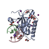
| Structure viewer | Molecule: MolmilJmol/JSmol |
|---|
- Downloads & links
-Download
| PDBx/mmCIF format | 2vog.cif.gz | 48 KB | Display | PDBx/mmCIF format |
|---|---|---|---|---|
| PDB format | pdb2vog.ent.gz | 33.2 KB | Display | PDB format |
| PDBx/mmJSON format | 2vog.json.gz | Tree view | PDBx/mmJSON format | |
| Others |  Other downloads Other downloads |
-Validation report
| Summary document | 2vog_validation.pdf.gz | 408.3 KB | Display | wwPDB validaton report |
|---|---|---|---|---|
| Full document | 2vog_full_validation.pdf.gz | 408.8 KB | Display | |
| Data in XML | 2vog_validation.xml.gz | 5.5 KB | Display | |
| Data in CIF | 2vog_validation.cif.gz | 7.4 KB | Display | |
| Arichive directory | https://data.pdbj.org/pub/pdb/validation_reports/vo/2vogftp://data.pdbj.org/pub/pdb/validation_reports/vo/2vog | HTTPS FTP |
-Related structure data
| Related structure data |  2vofSC 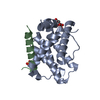 2vohC 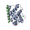 2voiC S: Starting model for refinement C: citing same article ( |
|---|---|
| Similar structure data |

-Links
 PDBj
PDBj
- Assembly
| Deposited unit | 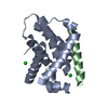
| ||||||||
|---|---|---|---|---|---|---|---|---|---|
| 1 |
| ||||||||
| Unit cell |
| ||||||||
| Components on special symmetry positions |
|
-Components
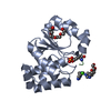
| #1: Protein | Mass: 17922.418 Da / Num. of mol.: 1 / Fragment: RESIDUES 1-152 / Mutation: YES Source method: isolated from a genetically manipulated source Source: (gene. exp.)  | ||||
|---|---|---|---|---|---|
| #2: Protein/peptide | Mass: 3275.765 Da / Num. of mol.: 1 / Fragment: BH3-DOMAIN, RESIDUES 126-152 Source method: isolated from a genetically manipulated source Details: C-TERMINAL HOMOSERINE LACTONE DUE TO CNBR CLEAVAGE. Source: (gene. exp.) | ||||
| #3: Chemical |   Mass: 35.453 Da / Num. of mol.: 2 / Source method: obtained synthetically / Formula: Cl Mass: 35.453 Da / Num. of mol.: 2 / Source method: obtained synthetically / Formula: Cl#4: Water | ChemComp-HOH / |  Mass: 18.015 Da / Num. of mol.: 49 / Source method: isolated from a natural source / Formula: H2O Mass: 18.015 Da / Num. of mol.: 49 / Source method: isolated from a natural source / Formula: H2OCompound details | ENGINEERED | |
-Experimental details
-Experiment
| Experiment | Method: X-RAY DIFFRACTION / Number of used crystals: 1 |
|---|
- Sample preparation
| Crystal | Density Matthews: 1.72 Å3/Da / Density % sol: 29 % / Description: NONE |
|---|---|
| Crystal grow | Details: 18% PEG 2000, 0.88 M LICL, 0.1 M CITRIC ACID, KOH (PH 4.35) |
-Data collection
| Diffraction | Mean temperature: 93 K |
|---|---|
| Diffraction source | Source: ROTATING ANODE / Type: RIGAKU MICROMAX-007 HF / Wavelength: 1.5418 |
| Detector | Type: RIGAKU IMAGE PLATE / Detector: IMAGE PLATE |
| Radiation | Protocol: SINGLE WAVELENGTH / Monochromatic (M) / Laue (L): M / Scattering type: x-ray |
| Radiation wavelength | Wavelength: 1.5418 Å / Relative weight: 1 |
| Reflection | Resolution: 1.9→23.81 Å / Num. obs: 12089 / % possible obs: 99.9 % / Observed criterion σ(I): 1 / Redundancy: 4 % / Rmerge(I) obs: 0.06 / Net I/σ(I): 19.1 |
| Reflection shell | Resolution: 1.9→2 Å / Redundancy: 3.7 % / Rmerge(I) obs: 0.36 / Mean I/σ(I) obs: 2.9 / % possible all: 99.8 |
- Processing
| Software |
| ||||||||||||||||||||||||||||||||||||||||||||||||||||||||||||||||||||||||||||||||||||||||||||||||||||||||||||||||||||||||||||||||||||||||||||||||||||||||||||||||||||||||||||||||||||||
|---|---|---|---|---|---|---|---|---|---|---|---|---|---|---|---|---|---|---|---|---|---|---|---|---|---|---|---|---|---|---|---|---|---|---|---|---|---|---|---|---|---|---|---|---|---|---|---|---|---|---|---|---|---|---|---|---|---|---|---|---|---|---|---|---|---|---|---|---|---|---|---|---|---|---|---|---|---|---|---|---|---|---|---|---|---|---|---|---|---|---|---|---|---|---|---|---|---|---|---|---|---|---|---|---|---|---|---|---|---|---|---|---|---|---|---|---|---|---|---|---|---|---|---|---|---|---|---|---|---|---|---|---|---|---|---|---|---|---|---|---|---|---|---|---|---|---|---|---|---|---|---|---|---|---|---|---|---|---|---|---|---|---|---|---|---|---|---|---|---|---|---|---|---|---|---|---|---|---|---|---|---|---|---|
| Refinement | Method to determine structure: MOLECULAR REPLACEMENT Starting model: PDB ENTRY 2VOF Resolution: 1.9→23.81 Å / Cor.coef. Fo:Fc: 0.95 / Cor.coef. Fo:Fc free: 0.939 / SU B: 6.251 / SU ML: 0.104 / TLS residual ADP flag: LIKELY RESIDUAL / Cross valid method: THROUGHOUT / ESU R: 0.179 / ESU R Free: 0.155 / Stereochemistry target values: MAXIMUM LIKELIHOOD / Details: HYDROGENS HAVE BEEN ADDED IN THE RIDING POSITIONS.
| ||||||||||||||||||||||||||||||||||||||||||||||||||||||||||||||||||||||||||||||||||||||||||||||||||||||||||||||||||||||||||||||||||||||||||||||||||||||||||||||||||||||||||||||||||||||
| Solvent computation | Ion probe radii: 0.8 Å / Shrinkage radii: 0.8 Å / VDW probe radii: 1.2 Å / Solvent model: BABINET MODEL WITH MASK | ||||||||||||||||||||||||||||||||||||||||||||||||||||||||||||||||||||||||||||||||||||||||||||||||||||||||||||||||||||||||||||||||||||||||||||||||||||||||||||||||||||||||||||||||||||||
| Displacement parameters | Biso mean: 26.59 Å2
| ||||||||||||||||||||||||||||||||||||||||||||||||||||||||||||||||||||||||||||||||||||||||||||||||||||||||||||||||||||||||||||||||||||||||||||||||||||||||||||||||||||||||||||||||||||||
| Refinement step | Cycle: LAST / Resolution: 1.9→23.81 Å
| ||||||||||||||||||||||||||||||||||||||||||||||||||||||||||||||||||||||||||||||||||||||||||||||||||||||||||||||||||||||||||||||||||||||||||||||||||||||||||||||||||||||||||||||||||||||
| Refine LS restraints |
|