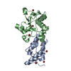
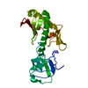
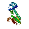
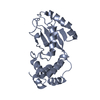
sporulation resulting in formation of a cellular spore / phosphorelay sensor kinase activity / histidine kinase / regulation of DNA-templated transcription / signal transduction / ATP binding / identical protein binding Similarity search - Function
PAS fold-3 / PAS fold / His Kinase A (phospho-acceptor) domain / His Kinase A (phosphoacceptor) domain / Signal transduction histidine kinase, dimerisation/phosphoacceptor domain / PAC motif / Motif C-terminal to PAS motifs (likely to contribute to PAS structural domain) / PAS domain / Signal transduction histidine kinase-related protein, C-terminal / Signal transduction histidine kinase, dimerisation/phosphoacceptor domain superfamily ...PAS fold-3 / PAS fold / His Kinase A (phospho-acceptor) domain / His Kinase A (phosphoacceptor) domain / Signal transduction histidine kinase, dimerisation/phosphoacceptor domain / PAC motif / Motif C-terminal to PAS motifs (likely to contribute to PAS structural domain) / PAS domain / Signal transduction histidine kinase-related protein, C-terminal / Signal transduction histidine kinase, dimerisation/phosphoacceptor domain superfamily / Histidine kinase domain / Histidine kinase domain profile. / Beta-Lactamase / PAS fold / PAS fold / PAS domain / PAS repeat profile. / Histidine kinase-, DNA gyrase B-, and HSP90-like ATPase / PAS domain / PAS domain superfamily / Histidine kinase-like ATPases / Histidine kinase/HSP90-like ATPase / Histidine kinase/HSP90-like ATPase superfamily / 2-Layer Sandwich / Alpha Beta Similarity search - Domain/homology
Mass: 18.015 Da / Num. of mol.: 301 / Source method: isolated from a natural source / Formula: H2O
Sequence details
RESIDUES 7-9 (GEF) BELONG TO THE CLONING LINKER
-
Experimental details
-
Experiment
Experiment
Method: X-RAY DIFFRACTION / Number of used crystals: 1
-
Sample preparation
Crystal
Density Matthews: 2.1 Å3/Da / Density % sol: 42.4 % / Description: NONE
Crystal grow
Temperature: 293 K / Method: vapor diffusion, hanging drop / pH: 5.5 Details: 20 DEGREES CELSIUS, HANGING-DROP VAPOR DIFFUSION: 1 MICROLITER KINA (7 MG/ML IN 25 MM TRIS, PH 8, 100 MM NACL PLUS 1 MICROLITER WELL SOLUTION (13 TO 15% (W/V) PEG 10,000, 0.1 M AMMONIUM ...Details: 20 DEGREES CELSIUS, HANGING-DROP VAPOR DIFFUSION: 1 MICROLITER KINA (7 MG/ML IN 25 MM TRIS, PH 8, 100 MM NACL PLUS 1 MICROLITER WELL SOLUTION (13 TO 15% (W/V) PEG 10,000, 0.1 M AMMONIUM ACETATE, 0.1 M BIS-TRIS, PH 5.5); CRYSTALS APPEARED AFTER ABOUT ONE DAY AND GREW TO A FINAL SIZE OF ABOUT 80 X 80 X 100 MICROMETERS WITHIN FOUR DAYS; CRYO-PROTECTION: STEPWISE TRANSFER INTO WELL SOLUTION SUPPLEMENTED WITH UP TO 25% (V/V) GLYCEROL.
Protocol: SINGLE WAVELENGTH / Monochromatic (M) / Laue (L): M / Scattering type: x-ray
Radiation wavelength
Wavelength: 0.97924 Å / Relative weight: 1
Reflection
Resolution: 1.71→44.75 Å / Num. obs: 42220 / % possible obs: 88 % / Observed criterion σ(I): -3 / Redundancy: 2.9 % / Biso Wilson estimate: 24.8 Å2 / Rmerge(I) obs: 0.04 / Net I/σ(I): 24.9
Reflection shell
Resolution: 1.71→1.74 Å / Redundancy: 2.4 % / Rmerge(I) obs: 0.42 / Mean I/σ(I) obs: 1.9 / % possible all: 56
-
Processing
Software
Name
Version
Classification
REFMAC
5.3.0037
refinement
HKL-2000
datareduction
HKL-2000
datascaling
SHELX
phasing
MLPHARE
phasing
DM
phasing
Refinement
Method to determine structure: SAD Starting model: NONE Resolution: 1.7→44 Å / Cor.coef. Fo:Fc: 0.965 / Cor.coef. Fo:Fc free: 0.947 / SU B: 5.938 / SU ML: 0.097 / Cross valid method: THROUGHOUT / ESU R: 0.13 / ESU R Free: 0.129 / Stereochemistry target values: MAXIMUM LIKELIHOOD / Details: HYDROGENS HAVE BEEN ADDED IN THE RIDING POSITIONS.
Rfactor
Num. reflection
% reflection
Selection details
Rfree
0.243
1499
3.5 %
RANDOM
Rwork
0.198
-
-
-
obs
0.2
40811
87.9 %
-
Solvent computation
Ion probe radii: 0.8 Å / Shrinkage radii: 0.8 Å / VDW probe radii: 1.2 Å / Solvent model: MASK
 Movie
Movie Controller


 Open data
 Basic information
 Components
 Keywords
 Function and homology information
 X-RAY DIFFRACTION /
  Authors
 Citation
 Structure visualization
 Downloads & links
 Other downloads
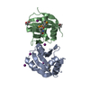


 PDBj
PDBj


 Assembly


 Mass: 35.453 Da / Num. of mol.: 4 / Source method: obtained synthetically / Formula: Cl
 Mass: 59.044 Da / Num. of mol.: 2 / Source method: obtained synthetically / Formula: C2H3O2
 Mass: 18.015 Da / Num. of mol.: 301 / Source method: isolated from a natural source / Formula: H2O
 Sample preparation
 / Beamline: 19-ID / Wavelength: 0.97924
  Processing