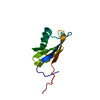
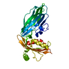
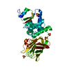
- PDB-2vj0: Crystal structure of the alpha-adaptin appendage domain, from the... -
+
Open data
ID or keywords:
Loading...
-
Basic information
Entry
Database: PDB / ID: 2vj0
Title
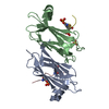
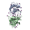
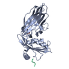
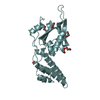
Crystal structure of the alpha-adaptin appendage domain, from the AP2 adaptor complex, in complex with an FXDNF peptide from amphiphysin1 and a WVXF peptide from synaptojanin P170
SHEET THE SHEET STRUCTURE OF THIS MOLECULE IS BIFURCATED. IN ORDER TO REPRESENT THIS FEATURE IN ... SHEET THE SHEET STRUCTURE OF THIS MOLECULE IS BIFURCATED. IN ORDER TO REPRESENT THIS FEATURE IN THE SHEET RECORDS BELOW, TWO SHEETS ARE DEFINED.
Mass: 18.015 Da / Num. of mol.: 343 / Source method: isolated from a natural source / Formula: H2O
-
Experimental details
-
Experiment
Experiment
Method: X-RAY DIFFRACTION / Number of used crystals: 1
-
Sample preparation
Crystal
Density Matthews: 3.2 Å3/Da / Density % sol: 61.1 % Description: THE ORIGINAL STRUCTURE BOUND ONLY TO THE SYNAPTOJANIN-1 P170 PEPTIDE, NOT REPORTED IN THE PAPER, WAS SOLVED BY MOLECULAR REPLACEMENT. A DIFFERENCE FOURIER WAS CALCULATED FOR THE SOAK OF ...Description: THE ORIGINAL STRUCTURE BOUND ONLY TO THE SYNAPTOJANIN-1 P170 PEPTIDE, NOT REPORTED IN THE PAPER, WAS SOLVED BY MOLECULAR REPLACEMENT. A DIFFERENCE FOURIER WAS CALCULATED FOR THE SOAK OF THE AMPHIPHYSIN PEPTIDE.
Resolution: 1.6→17.76 Å / Cor.coef. Fo:Fc: 0.958 / Cor.coef. Fo:Fc free: 0.944 / SU B: 1.42 / SU ML: 0.051 / Cross valid method: THROUGHOUT / ESU R: 0.079 / ESU R Free: 0.08 / Stereochemistry target values: MAXIMUM LIKELIHOOD / Details: HYDROGENS HAVE BEEN ADDED IN THE RIDING POSITIONS.
Rfactor
Num. reflection
% reflection
Selection details
Rfree
0.208
2427
5.18 %
RANDOM
Rwork
0.181
-
-
-
obs
0.182
47892
96.2 %
-
Solvent computation
Ion probe radii: 0.8 Å / VDW probe radii: 1.4 Å / Solvent model: BABINET MODEL PLUS MASK
 Movie
Movie Controller

 Yorodumi
 Open data
 Basic information
 Components
 Keywords
 Function and homology information
 HOMO SAPIENS (human)
 X-RAY DIFFRACTION /
  Authors
 Citation
 Structure visualization
 Downloads & links
 Other downloads


 PDBj
PDBj


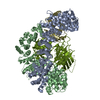
 Assembly


 Mass: 120.152 Da / Num. of mol.: 1 / Source method: obtained synthetically / Formula: C7H8N2
 Mass: 152.235 Da / Num. of mol.: 1 / Source method: obtained synthetically / Formula: C4H8O2S2
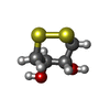
 Mass: 96.063 Da / Num. of mol.: 3 / Source method: obtained synthetically / Formula: SO4
 Mass: 35.453 Da / Num. of mol.: 1 / Source method: obtained synthetically / Formula: Cl
 Sample preparation
 / Beamline: PX9.6 / Wavelength: 0.87
  Processing