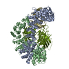
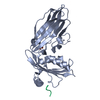
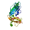
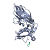
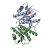
SHEET THE SHEET STRUCTURE OF THIS MOLECULE IS BIFURCATED. IN ORDER TO REPRESENT THIS FEATURE IN ... SHEET THE SHEET STRUCTURE OF THIS MOLECULE IS BIFURCATED. IN ORDER TO REPRESENT THIS FEATURE IN THE SHEET RECORDS BELOW, TWO SHEETS ARE DEFINED.
Mass: 18.015 Da / Num. of mol.: 277 / Source method: isolated from a natural source / Formula: H2O
-
Details
Compound details
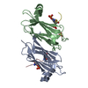
CATALYSES THE FOLLOWING REACTION: 1-PHOSPHATIDYL-1D-MYO-INOSITOL 4,5-BISPHOSPHATE + H2O --> 1- ...CATALYSES THE FOLLOWING REACTION: 1-PHOSPHATIDYL-1D-MYO-INOSITOL 4,5-BISPHOSPHATE + H2O --> 1-PHOSPHATIDYL-1D-MYO-INOSITOL 4-PHOSPHATE + PHOSPHATE.
Sequence details
AT RESIDUES 889 AND 890 FOR CHAIN A, THE SEQUENCE IN THE COORDINATES DISAGREES WITH THE UNIPROT ...AT RESIDUES 889 AND 890 FOR CHAIN A, THE SEQUENCE IN THE COORDINATES DISAGREES WITH THE UNIPROT ENTRY. THE SEQUENCE IN THE COORDINATES (GLY-ALA) AGREES WITH THE ELECTRON DENSITY AND SEQUENCING EXPERIMENTS. THE FIRST THREE RESIDUES FOR WHICH DENSITY IS OBSERVED FOR CHAIN A (PRO-GLY-ILE) ARE FROM THE EXPRESSION VECTOR (PGEX-4T2). THIS IS PRECEEDED BY GLY-SER FOR WHICH NO DENSITY IS OBSERVED. PRO-ILE-GLY IS FOLLOWED BY A LEU WHICH WAS INTRODUCED IN THE CLONING PRIMER. THE SEQUENCE FOLLOWING THIS RESIDUE MATCHES UNIPROT REFERENCE P17427.
-
Experimental details
-
Experiment
Experiment
Method: X-RAY DIFFRACTION / Number of used crystals: 1
-
Sample preparation
Crystal
Density Matthews: 3.6 Å3/Da / Density % sol: 65.9 %
Resolution: 1.9→72.55 Å / Cor.coef. Fo:Fc: 0.965 / Cor.coef. Fo:Fc free: 0.941 / SU B: 3.451 / SU ML: 0.1 / Cross valid method: THROUGHOUT / ESU R: 0.13 / ESU R Free: 0.132 / Stereochemistry target values: MAXIMUM LIKELIHOOD / Details: HYDROGENS HAVE BEEN ADDED IN THE RIDING POSITIONS.
Rfactor
Num. reflection
% reflection
Selection details
Rfree
0.224
1467
5.1 %
RANDOM
Rwork
0.177
-
-
-
obs
0.179
27464
95 %
-
Solvent computation
Ion probe radii: 0.8 Å / Shrinkage radii: 0.8 Å / VDW probe radii: 1.2 Å / Solvent model: BABINET MODEL WITH MASK
 Movie
Movie Controller

 Yorodumi
 Open data
 Basic information
 Components
 Keywords
 Function and homology information
 HOMO SAPIENS (human)
 X-RAY DIFFRACTION /
  Authors
 Citation
 Structure visualization
 Downloads & links
 Other downloads


 PDBj
PDBj


 Assembly


 Mass: 120.152 Da / Num. of mol.: 1 / Source method: obtained synthetically / Formula: C7H8N2
 Mass: 60.009 Da / Num. of mol.: 3 / Source method: obtained synthetically / Formula: CO3
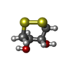
 Mass: 152.235 Da / Num. of mol.: 1 / Source method: obtained synthetically / Formula: C4H8O2S2
 Mass: 96.063 Da / Num. of mol.: 1 / Source method: obtained synthetically / Formula: SO4
 Sample preparation
 / Beamline: ID14-1 / Wavelength: 0.98
  Processing