 Movie
Movie Controller

[English] 日本語
 Yorodumi
- PDB-2vi7: Structure of a Putative Acetyltransferase (PA1377)from Pseudomona... -
+ Open data

- Basic information
| Entry | Database: PDB / ID: 2vi7 | ||||||
|---|---|---|---|---|---|---|---|
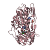
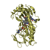
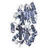
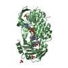
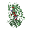
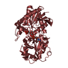
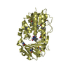
| Title | Structure of a Putative Acetyltransferase (PA1377)from Pseudomonas aeruginosa | ||||||
 Components Components | ACETYLTRANSFERASE PA1377 | ||||||
 Keywords Keywords | TRANSFERASE / GNAT / GCN5 FAMILY / N-ACETYLTRANSFERASE / HYPOTHETICAL PROTEIN | ||||||
| Function / homology |  Function and homology information Function and homology information | ||||||
| Biological species |   PSEUDOMONAS AERUGINOSA (bacteria) PSEUDOMONAS AERUGINOSA (bacteria) | ||||||
| Method |  X-RAY DIFFRACTION / SYNCHROTRON / MOLECULAR REPLACEMENT / Resolution: 2.25 Å X-RAY DIFFRACTION / SYNCHROTRON / MOLECULAR REPLACEMENT / Resolution: 2.25 Å | ||||||
 Authors Authors | Davies, A.M. / Tata, R. / Chauviac, F.X. / Sutton, B.J. / Brown, P.R. | ||||||
 Citation Citation | Journal: Acta Crystallogr.,Sect.F / Year: 2008 Title: Structure of a Putative Acetyltransferase (Pa1377) from Pseudomonas Aeruginosa. Authors: Davies, A.M. / Tata, R. / Chauviac, F.X. / Sutton, B.J. / Brown, P.R. | ||||||
| History |
| ||||||
| Remark 700 | SHEET THE SHEET STRUCTURE OF THIS MOLECULE IS BIFURCATED. IN ORDER TO REPRESENT THIS FEATURE IN ... SHEET THE SHEET STRUCTURE OF THIS MOLECULE IS BIFURCATED. IN ORDER TO REPRESENT THIS FEATURE IN THE SHEET RECORDS BELOW, TWO SHEETS ARE DEFINED. |
- Structure visualization
| Structure viewer | Molecule: MolmilJmol/JSmol |
|---|
- Downloads & links
-Download
| PDBx/mmCIF format | 2vi7.cif.gz | 115.5 KB | Display | PDBx/mmCIF format |
|---|---|---|---|---|
| PDB format | pdb2vi7.ent.gz | 89.1 KB | Display | PDB format |
| PDBx/mmJSON format | 2vi7.json.gz | Tree view | PDBx/mmJSON format | |
| Others |  Other downloads Other downloads |
-Validation report
| Arichive directory | https://data.pdbj.org/pub/pdb/validation_reports/vi/2vi7ftp://data.pdbj.org/pub/pdb/validation_reports/vi/2vi7 | HTTPS FTP |
|---|
-Related structure data
| Related structure data |  2ge3S S: Starting model for refinement |
|---|---|
| Similar structure data |


-Links
 PDBj
PDBj

- Assembly
| Deposited unit | 
| ||||||||||||
|---|---|---|---|---|---|---|---|---|---|---|---|---|---|
| 1 | 
| ||||||||||||
| 2 | 
| ||||||||||||
| Unit cell |
| ||||||||||||
| Components on special symmetry positions |
| ||||||||||||
| Noncrystallographic symmetry (NCS) | NCS oper:
|
-Components
| #1: Protein | Mass: 20097.773 Da / Num. of mol.: 3 Source method: isolated from a genetically manipulated source Source: (gene. exp.) PSEUDOMONAS AERUGINOSA (bacteria) / Strain: PAC1 / Plasmid: PET24A / Production host: #2: Chemical | ChemComp-GOL /   Mass: 92.094 Da / Num. of mol.: 5 / Source method: obtained synthetically / Formula: C3H8O3 Mass: 92.094 Da / Num. of mol.: 5 / Source method: obtained synthetically / Formula: C3H8O3#3: Chemical | ChemComp-SO4 /   Mass: 96.063 Da / Num. of mol.: 6 / Source method: obtained synthetically / Formula: SO4 Mass: 96.063 Da / Num. of mol.: 6 / Source method: obtained synthetically / Formula: SO4#4: Chemical | ChemComp-AZI /   Mass: 42.020 Da / Num. of mol.: 4 / Source method: obtained synthetically / Formula: N3 Mass: 42.020 Da / Num. of mol.: 4 / Source method: obtained synthetically / Formula: N3#5: Water | ChemComp-HOH / |  Mass: 18.015 Da / Num. of mol.: 265 / Source method: isolated from a natural source / Formula: H2O Mass: 18.015 Da / Num. of mol.: 265 / Source method: isolated from a natural source / Formula: H2O |
|---|
-Experimental details
-Experiment
| Experiment | Method: X-RAY DIFFRACTION / Number of used crystals: 1 |
|---|
- Sample preparation
| Crystal | Density Matthews: 2.48 Å3/Da / Density % sol: 50.4 % / Description: NONE |
|---|---|
| Crystal grow | Temperature: 291 K / Method: vapor diffusion, hanging drop / pH: 6.5 Details: HANGING DROP VAPOR DIFFUSION. RESERVOIR OF 500 MICROLITRES 0.1M TRIS-HCL AT PH6.5, 1.6M AMMONIUM SULFATE AND 0.1% SODIUM AZIDE. DROPS CONTAINED 1 MICROLITRE OF PROTEIN AT 13MG /ML AND AN ...Details: HANGING DROP VAPOR DIFFUSION. RESERVOIR OF 500 MICROLITRES 0.1M TRIS-HCL AT PH6.5, 1.6M AMMONIUM SULFATE AND 0.1% SODIUM AZIDE. DROPS CONTAINED 1 MICROLITRE OF PROTEIN AT 13MG /ML AND AN EQUAL VOLUME OF RESERVOIR. DROPS WERE KEPT AT 291K. |
-Data collection
| Diffraction | Mean temperature: 100 K |
|---|---|
| Diffraction source | Source: SYNCHROTRON / Site: SRS  / Beamline: PX10.1 / Wavelength: 1.488 / Beamline: PX10.1 / Wavelength: 1.488 |
| Detector | Type: MARRESEARCH / Detector: CCD / Date: Apr 19, 2005 |
| Radiation | Protocol: SINGLE WAVELENGTH / Monochromatic (M) / Laue (L): M / Scattering type: x-ray |
| Radiation wavelength | Wavelength: 1.488 Å / Relative weight: 1 |
| Reflection | Resolution: 2.25→64.3 Å / Num. obs: 28683 / % possible obs: 99.9 % / Observed criterion σ(I): 0 / Redundancy: 6.7 % / Biso Wilson estimate: 39.5 Å2 / Rmerge(I) obs: 0.09 / Net I/σ(I): 21 |
| Reflection shell | Resolution: 2.25→2.31 Å / Redundancy: 6.6 % / Rmerge(I) obs: 0.61 / Mean I/σ(I) obs: 2.8 / % possible all: 99.9 |
- Processing
| Software |
| ||||||||||||||||||||||||||||||||||||||||||||||||||||||||||||
|---|---|---|---|---|---|---|---|---|---|---|---|---|---|---|---|---|---|---|---|---|---|---|---|---|---|---|---|---|---|---|---|---|---|---|---|---|---|---|---|---|---|---|---|---|---|---|---|---|---|---|---|---|---|---|---|---|---|---|---|---|---|
| Refinement | Method to determine structure: MOLECULAR REPLACEMENT Starting model: PDB ENTRY 2GE3 Resolution: 2.25→64.3 Å / Cross valid method: THROUGHOUT / σ(F): 0 / Stereochemistry target values: MAXIMUM LIKELIHOOD Details: MOLECULES A AND B OF THE ASYMMETRIC UNIT FORM THE ASSUMED DIMERIC, BIOLOGICAL UNIT. MOLECULE C FORMS A DIMER WITH A MOLECULE RELATED BY CRYSTALLOGRAPHIC SYMMETRY.
| ||||||||||||||||||||||||||||||||||||||||||||||||||||||||||||
| Solvent computation | Bsol: 42.52 Å2 / ksol: 0.35 e/Å3 | ||||||||||||||||||||||||||||||||||||||||||||||||||||||||||||
| Displacement parameters | Biso mean: 37 Å2 | ||||||||||||||||||||||||||||||||||||||||||||||||||||||||||||
| Refine analyze |
| ||||||||||||||||||||||||||||||||||||||||||||||||||||||||||||
| Refinement step | Cycle: LAST / Resolution: 2.25→64.3 Å
| ||||||||||||||||||||||||||||||||||||||||||||||||||||||||||||
| Refine LS restraints |
| ||||||||||||||||||||||||||||||||||||||||||||||||||||||||||||
| LS refinement shell | Resolution: 2.25→2.33 Å / Num. reflection Rfree: 136 / Num. reflection Rwork: 2684 / Total num. of bins used: 10 |
