SHEET THE SHEET STRUCTURE OF THIS MOLECULE IS BIFURCATED. IN ORDER TO REPRESENT THIS FEATURE IN ... SHEET THE SHEET STRUCTURE OF THIS MOLECULE IS BIFURCATED. IN ORDER TO REPRESENT THIS FEATURE IN THE SHEET RECORDS BELOW, TWO SHEETS ARE DEFINED.
Mass: 18.015 Da / Num. of mol.: 590 / Source method: isolated from a natural source / Formula: H2O
Compound details
ENGINEERED RESIDUE IN CHAIN A, SER 252 TO ALA ENGINEERED RESIDUE IN CHAIN B, SER 252 TO ALA ...ENGINEERED RESIDUE IN CHAIN A, SER 252 TO ALA ENGINEERED RESIDUE IN CHAIN B, SER 252 TO ALA ENGINEERED RESIDUE IN CHAIN C, SER 252 TO ALA
Sequence details
POINT MUTATION INTRODUCED AT S252 INTO A252 WHEN COMPARED TO UNIPROT Q94M05
-
Experimental details
-
Experiment
Experiment
Method: X-RAY DIFFRACTION / Number of used crystals: 1
-
Sample preparation
Crystal
Density Matthews: 2.6 Å3/Da / Density % sol: 52 % Description: CRYSTALS OF P4 MUTANT S252A IN COMPLEX WITH ATP AND MG ARE ISOMORPOUS WITH P4-AMPCPP-MG CRYSTALS
Monochromator: SI(111) / Protocol: SINGLE WAVELENGTH / Monochromatic (M) / Laue (L): M / Scattering type: x-ray
Radiation wavelength
Wavelength: 1.072 Å / Relative weight: 1
Reflection
Resolution: 2.15→30 Å / Num. obs: 98926 / % possible obs: 100 % / Observed criterion σ(I): 2 / Redundancy: 7.8 % / Rmerge(I) obs: 0.13 / Net I/σ(I): 15
Reflection shell
Resolution: 2.15→2.23 Å / Redundancy: 7.5 % / Rmerge(I) obs: 0.87 / Mean I/σ(I) obs: 1.8 / % possible all: 100
-
Processing
Software
Name
Version
Classification
REFMAC
5.2.0019
refinement
DENZO
datareduction
SCALEPACK
datascaling
CNS
phasing
Refinement
Method to determine structure: OTHER Starting model: NONE Resolution: 2.15→100.5 Å / Cor.coef. Fo:Fc: 0.947 / Cor.coef. Fo:Fc free: 0.918 / SU B: 4.85 / SU ML: 0.131 / Cross valid method: THROUGHOUT / ESU R: 0.23 / ESU R Free: 0.198 / Stereochemistry target values: MAXIMUM LIKELIHOOD / Details: HYDROGENS HAVE BEEN ADDED IN THE RIDING POSITIONS.
Rfactor
Num. reflection
% reflection
Selection details
Rfree
0.247
2994
5.1 %
RANDOM
Rwork
0.195
-
-
-
obs
0.198
56185
99.9 %
-
Solvent computation
Ion probe radii: 0.8 Å / Shrinkage radii: 0.8 Å / VDW probe radii: 1.4 Å / Solvent model: BABINET MODEL WITH MASK
 Movie
Movie Controller

 Yorodumi
 Open data
 Basic information
 Components
 Keywords
 Function and homology information
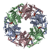
 PSEUDOMONAS PHAGE PHI12 (virus)
 X-RAY DIFFRACTION /
  Authors
 Citation
 Structure visualization
 Downloads & links
 Other downloads


 PDBj
PDBj Assembly
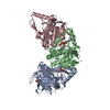
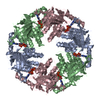


 Mass: 507.181 Da / Num. of mol.: 3 / Source method: obtained synthetically / Formula: C10H16N5O13P3 / Comment: ATP, energy-carrying molecule*YM
 Mass: 24.305 Da / Num. of mol.: 3 / Source method: obtained synthetically / Formula: Mg
 Mass: 18.015 Da / Num. of mol.: 590 / Source method: isolated from a natural source / Formula: H2O
 Sample preparation
 / Beamline: ID23-1 / Wavelength: 1.072
  Processing