 Movie
Movie Controller

[English] 日本語
 Yorodumi
- PDB-2ves: Crystal Structure of LpxC from Pseudomonas aeruginosa complexed w... -
+ Open data

- Basic information
| Entry | Database: PDB / ID: 2ves | ||||||
|---|---|---|---|---|---|---|---|
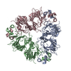
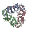
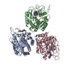
| Title | Crystal Structure of LpxC from Pseudomonas aeruginosa complexed with the potent BB-78485 inhibitor | ||||||
 Components Components | UDP-3-O-acyl-N-acetylglucosamine deacetylase | ||||||
 Keywords Keywords | HYDROLASE / LPXC / BB-78485 / ANTIBIOTICS / DEACETYLASE / LIPID SYNTHESIS / METALLOPROTEASE / HYDROXAMIC ACID / GRAM-NEGATIVE BACTERIA / LIPOPOLYSACCHARIDE / LIPID A BIOSYNTHESIS | ||||||
| Function / homology |  Function and homology information Function and homology informationUDP-3-O-acyl-N-acetylglucosamine deacetylase / UDP-3-O-acyl-N-acetylglucosamine deacetylase activity / lipid A biosynthetic process / membrane / metal ion binding / cytoplasm Similarity search - Function | ||||||
| Biological species |   Pseudomonas aeruginosa (bacteria) Pseudomonas aeruginosa (bacteria) | ||||||
| Method |  X-RAY DIFFRACTION / SYNCHROTRON / MOLECULAR REPLACEMENT / Resolution: 1.9 Å X-RAY DIFFRACTION / SYNCHROTRON / MOLECULAR REPLACEMENT / Resolution: 1.9 Å | ||||||
 Authors Authors | Mochalkin, I. / Knafels, J.D. | ||||||
 Citation Citation | Journal: Protein Sci. / Year: 2008 Title: Crystal structure of LpxC from Pseudomonas aeruginosa complexed with the potent BB-78485 inhibitor. Authors: Mochalkin, I. / Knafels, J.D. / Lightle, S. | ||||||
| History |
| ||||||
| Remark 700 | SHEET THE SHEET STRUCTURE OF THIS MOLECULE IS BIFURCATED. IN ORDER TO REPRESENT THIS FEATURE IN ... SHEET THE SHEET STRUCTURE OF THIS MOLECULE IS BIFURCATED. IN ORDER TO REPRESENT THIS FEATURE IN THE SHEET RECORDS BELOW, TWO SHEETS ARE DEFINED. |
- Structure visualization
| Structure viewer | Molecule: MolmilJmol/JSmol |
|---|
- Downloads & links
-Download
| PDBx/mmCIF format | 2ves.cif.gz | 211.6 KB | Display | PDBx/mmCIF format |
|---|---|---|---|---|
| PDB format | pdb2ves.ent.gz | 169.9 KB | Display | PDB format |
| PDBx/mmJSON format | 2ves.json.gz | Tree view | PDBx/mmJSON format | |
| Others |  Other downloads Other downloads |
-Validation report
| Arichive directory | https://data.pdbj.org/pub/pdb/validation_reports/ve/2vesftp://data.pdbj.org/pub/pdb/validation_reports/ve/2ves | HTTPS FTP |
|---|
-Related structure data
| Related structure data |  1p42S S: Starting model for refinement |
|---|---|
| Similar structure data |


-Links
 PDBj
PDBj- Assembly
| Deposited unit | 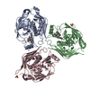
| ||||||||
|---|---|---|---|---|---|---|---|---|---|
| 1 |
| ||||||||
| Unit cell |
|
-Components
| #1: Protein | Mass: 33146.617 Da / Num. of mol.: 3 / Fragment: RESIDUES 1-299 / Mutation: C40S Source method: isolated from a genetically manipulated source Source: (gene. exp.) Pseudomonas aeruginosa (bacteria) / Strain: 287 / Gene: lpxC, envA, PA4406 / Production host: References: UniProt: P47205, UDP-3-O-acyl-N-acetylglucosamine deacetylase #2: Chemical | ChemComp-ZN /   Mass: 65.409 Da / Num. of mol.: 7 / Source method: obtained synthetically / Formula: Zn Mass: 65.409 Da / Num. of mol.: 7 / Source method: obtained synthetically / Formula: Zn#3: Chemical |   Mass: 420.481 Da / Num. of mol.: 3 / Source method: obtained synthetically / Formula: C23H20N2O4S Mass: 420.481 Da / Num. of mol.: 3 / Source method: obtained synthetically / Formula: C23H20N2O4S#4: Chemical |   Mass: 96.063 Da / Num. of mol.: 2 / Source method: obtained synthetically / Formula: SO4 Mass: 96.063 Da / Num. of mol.: 2 / Source method: obtained synthetically / Formula: SO4#5: Water | ChemComp-HOH / |  Mass: 18.015 Da / Num. of mol.: 1223 / Source method: isolated from a natural source / Formula: H2O Mass: 18.015 Da / Num. of mol.: 1223 / Source method: isolated from a natural source / Formula: H2OCompound details | ENGINEERED RESIDUE IN CHAIN A, CYS 40 TO SER ENGINEERED RESIDUE IN CHAIN B, CYS 40 TO SER ...ENGINEERED | Nonpolymer details | 3-PYRIDIN-4-YL-2,4-DIHYDRO-INDENO[1,2-.C.]PYRAZOLE (GVR): BB-78485 FROM BRITISH BIOTECH ...3-PYRIDIN-4-YL-2,4-DIHYDRO-INDENO[1,2-.C.]PYRAZOLE (GVR): BB-78485 FROM BRITISH BIOTECH PHARMACEUT | Sequence details | RESIDUES 1-299, C40S MUTATION. | |
|---|
-Experimental details
-Experiment
| Experiment | Method: X-RAY DIFFRACTION / Number of used crystals: 1 |
|---|
- Sample preparation
| Crystal | Density Matthews: 2.13 Å3/Da / Density % sol: 50.32 % / Description: NONE |
|---|---|
| Crystal grow | Method: vapor diffusion, hanging drop / pH: 6.5 Details: PALPXC, 10 MG/ML IN 25 MM HEPES PH 7.0, 2 MM TCEP, 0.3 M NACL, 1MM ZNCL2. BB-78485 ADDED TO 1 MM. HANGING DROP, 0.1 M SODIUM CACODYLATE PH 6.5, 0.2 M AMMONIUM SULFATE AND 10 % PEG 8000, 10 MM ZNCL2. |
-Data collection
| Diffraction | Mean temperature: 173 K |
|---|---|
| Diffraction source | Source: SYNCHROTRON / Site: APS  / Beamline: 17-ID / Wavelength: 1 / Beamline: 17-ID / Wavelength: 1 |
| Detector | Type: ADSC CCD / Detector: CCD / Date: Dec 1, 2004 / Details: MIRRORS |
| Radiation | Monochromator: NI FILTER / Protocol: SINGLE WAVELENGTH / Monochromatic (M) / Laue (L): M / Scattering type: x-ray |
| Radiation wavelength | Wavelength: 1 Å / Relative weight: 1 |
| Reflection | Resolution: 1.9→34.15 Å / Num. obs: 78112 / % possible obs: 99.9 % / Observed criterion σ(I): 0 / Redundancy: 6.4 % / Rmerge(I) obs: 0.08 / Net I/σ(I): 23.61 |
| Reflection shell | Resolution: 1.9→1.97 Å / Redundancy: 5.7 % / Rmerge(I) obs: 0.52 / Mean I/σ(I) obs: 2.5 / % possible all: 99.5 |
- Processing
| Software |
| ||||||||||||||||||||||||||||||||||||||||||||||||||||||||||||||||||||||||||||||||||||||||||||||||||||||||||||||||||||||||||||||||||||||||||||||||||||||||||||||||||||||||||||||||||||||
|---|---|---|---|---|---|---|---|---|---|---|---|---|---|---|---|---|---|---|---|---|---|---|---|---|---|---|---|---|---|---|---|---|---|---|---|---|---|---|---|---|---|---|---|---|---|---|---|---|---|---|---|---|---|---|---|---|---|---|---|---|---|---|---|---|---|---|---|---|---|---|---|---|---|---|---|---|---|---|---|---|---|---|---|---|---|---|---|---|---|---|---|---|---|---|---|---|---|---|---|---|---|---|---|---|---|---|---|---|---|---|---|---|---|---|---|---|---|---|---|---|---|---|---|---|---|---|---|---|---|---|---|---|---|---|---|---|---|---|---|---|---|---|---|---|---|---|---|---|---|---|---|---|---|---|---|---|---|---|---|---|---|---|---|---|---|---|---|---|---|---|---|---|---|---|---|---|---|---|---|---|---|---|---|
| Refinement | Method to determine structure: MOLECULAR REPLACEMENT Starting model: PDB ENTRY 1P42 Resolution: 1.9→73.72 Å / Cor.coef. Fo:Fc: 0.958 / Cor.coef. Fo:Fc free: 0.934 / SU B: 5.672 / SU ML: 0.094 / Cross valid method: THROUGHOUT / ESU R: 0.154 / ESU R Free: 0.143 / Stereochemistry target values: MAXIMUM LIKELIHOOD Details: HYDROGENS HAVE BEEN ADDED IN THE RIDING POSITIONS. THERE ARE THREE MOLECULES IN ASYMMETRIC UNIT - CHAINS A, B AND C. C-TERMINAL ENDS IN CHAIN A AND B ARE DISORDERED. CHAIN C IS COMPLETE.
| ||||||||||||||||||||||||||||||||||||||||||||||||||||||||||||||||||||||||||||||||||||||||||||||||||||||||||||||||||||||||||||||||||||||||||||||||||||||||||||||||||||||||||||||||||||||
| Solvent computation | Ion probe radii: 0.8 Å / Shrinkage radii: 0.8 Å / VDW probe radii: 1.4 Å / Solvent model: MASK | ||||||||||||||||||||||||||||||||||||||||||||||||||||||||||||||||||||||||||||||||||||||||||||||||||||||||||||||||||||||||||||||||||||||||||||||||||||||||||||||||||||||||||||||||||||||
| Displacement parameters | Biso mean: 23.05 Å2
| ||||||||||||||||||||||||||||||||||||||||||||||||||||||||||||||||||||||||||||||||||||||||||||||||||||||||||||||||||||||||||||||||||||||||||||||||||||||||||||||||||||||||||||||||||||||
| Refinement step | Cycle: LAST / Resolution: 1.9→73.72 Å
| ||||||||||||||||||||||||||||||||||||||||||||||||||||||||||||||||||||||||||||||||||||||||||||||||||||||||||||||||||||||||||||||||||||||||||||||||||||||||||||||||||||||||||||||||||||||
| Refine LS restraints |
|