 Movie
Movie Controller

[English] 日本語
 Yorodumi
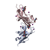
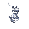
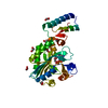
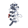
- PDB-2vdf: Structure of the OpcA adhesion from Neisseria meningitidis determ... -
+ Open data

- Basic information
| Entry | Database: PDB / ID: 2vdf | ||||||
|---|---|---|---|---|---|---|---|
| Title | Structure of the OpcA adhesion from Neisseria meningitidis determined by crystallization from the cubic mesophase | ||||||
 Components Components | OUTER MEMBRANE PROTEIN | ||||||
 Keywords Keywords | MEMBRANE PROTEIN / INVASIN / ADHESIN / BETA BARREL / OUTER MEMBRANE | ||||||
| Function / homology |  Function and homology information Function and homology information | ||||||
| Biological species |  NEISSERIA MENINGITIDIS (bacteria) NEISSERIA MENINGITIDIS (bacteria) | ||||||
| Method |  X-RAY DIFFRACTION / SYNCHROTRON / MOLECULAR REPLACEMENT / Resolution: 1.95 Å X-RAY DIFFRACTION / SYNCHROTRON / MOLECULAR REPLACEMENT / Resolution: 1.95 Å | ||||||
 Authors Authors | Cherezov, V. / Liu, W. / Derrick, J.P. / Luan, B. / Aksimentiev, A. / Katritch, V. / Caffrey, M. | ||||||
 Citation Citation | Journal: Proteins / Year: 2008 Title: In meso crystal structure and docking simulations suggest an alternative proteoglycan binding site in the OpcA outer membrane adhesin. Authors: Cherezov, V. / Liu, W. / Derrick, J.P. / Luan, B. / Aksimentiev, A. / Katritch, V. / Caffrey, M. #1: Journal: Proc.Natl.Acad.Sci.USA / Year: 2002Title: Crystal Structure of the Opca Integral Membrane Adhesin from Neisseria Meningitidis. Authors: Prince, S.M. / Achtman, M. / Derrick, J.P. | ||||||
| History |
| ||||||
| Remark 700 | SHEET DETERMINATION METHOD: DSSP THE SHEETS PRESENTED AS "AA" IN EACH CHAIN ON SHEET RECORDS BELOW ... SHEET DETERMINATION METHOD: DSSP THE SHEETS PRESENTED AS "AA" IN EACH CHAIN ON SHEET RECORDS BELOW IS ACTUALLY AN 10-STRANDED BARREL THIS IS REPRESENTED BY A 11-STRANDED SHEET IN WHICH THE FIRST AND LAST STRANDS ARE IDENTICAL. |
- Structure visualization
| Structure viewer | Molecule: MolmilJmol/JSmol |
|---|
- Downloads & links
-Download
| PDBx/mmCIF format | 2vdf.cif.gz | 106.6 KB | Display | PDBx/mmCIF format |
|---|---|---|---|---|
| PDB format | pdb2vdf.ent.gz | 80.6 KB | Display | PDB format |
| PDBx/mmJSON format | 2vdf.json.gz | Tree view | PDBx/mmJSON format | |
| Others |  Other downloads Other downloads |
-Validation report
| Arichive directory | https://data.pdbj.org/pub/pdb/validation_reports/vd/2vdfftp://data.pdbj.org/pub/pdb/validation_reports/vd/2vdf | HTTPS FTP |
|---|
-Related structure data
| Related structure data |  1k24S  2j9s S: Starting model for refinement |
|---|---|
| Similar structure data |


-Links
 PDBj
PDBj

- Assembly
| Deposited unit | 
| ||||||||
|---|---|---|---|---|---|---|---|---|---|
| 1 |
| ||||||||
| Unit cell |
|
-Components
| #1: Protein | Mass: 28092.643 Da / Num. of mol.: 1 Source method: isolated from a genetically manipulated source Source: (gene. exp.) NEISSERIA MENINGITIDIS (bacteria) / Production host: |
|---|---|
| #2: Chemical | ChemComp-OCT /   Mass: 114.229 Da / Num. of mol.: 1 / Source method: obtained synthetically / Formula: C8H18 Mass: 114.229 Da / Num. of mol.: 1 / Source method: obtained synthetically / Formula: C8H18 |
| #3: Chemical | ChemComp-SO4 /   Mass: 96.063 Da / Num. of mol.: 1 / Source method: obtained synthetically / Formula: SO4 Mass: 96.063 Da / Num. of mol.: 1 / Source method: obtained synthetically / Formula: SO4 |
| #4: Water | ChemComp-HOH /  Mass: 18.015 Da / Num. of mol.: 120 / Source method: isolated from a natural source / Formula: H2O Mass: 18.015 Da / Num. of mol.: 120 / Source method: isolated from a natural source / Formula: H2O |
| Nonpolymer details | SULFATE ION (SO4): FROM CRYSTALLIZATION CONDITIONS N-OCTANE (OCT): ORIGINATES FROM DETERGENT USED ...SULFATE ION (SO4): FROM CRYSTALLIZ |
-Experimental details
-Experiment
| Experiment | Method: X-RAY DIFFRACTION / Number of used crystals: 1 |
|---|
- Sample preparation
| Crystal | Density Matthews: 2.16 Å3/Da / Density % sol: 43 % / Description: NONE |
|---|---|
| Crystal grow | Temperature: 293 K / Method: lipidic cubic phase Details: 18%(V/V) PEG400, 0.1 M POTASSIUM SULFATE, 0.05 M HEPES, PH 7.0 |
-Data collection
| Diffraction | Mean temperature: 100 K |
|---|---|
| Diffraction source | Source: SYNCHROTRON / Site: CHESS  / Beamline: F1 / Wavelength: 0.9176 / Beamline: F1 / Wavelength: 0.9176 |
| Detector | Type: ADSC CCD / Detector: CCD |
| Radiation | Protocol: SINGLE WAVELENGTH / Monochromatic (M) / Laue (L): M / Scattering type: x-ray |
| Radiation wavelength | Wavelength: 0.9176 Å / Relative weight: 1 |
| Reflection | Resolution: 1.95→20 Å / Num. obs: 18131 / % possible obs: 98.8 % / Observed criterion σ(I): 2 / Redundancy: 4.8 % / Rmerge(I) obs: 0.099 / Net I/σ(I): 13.1 |
| Reflection shell | Resolution: 1.95→2.06 Å / Redundancy: 4.7 % / Rmerge(I) obs: 0.49 / Mean I/σ(I) obs: 2.7 / % possible all: 99.8 |
- Processing
| Software |
| ||||||||||||||||||||||||||||||||||||||||||||||||||||||||||||||||||||||||||||||||||||||||||||||||||||||||||||||||||||||||||||||||||||||||||||||||||||||||||||||||||||||||||||||||||||||
|---|---|---|---|---|---|---|---|---|---|---|---|---|---|---|---|---|---|---|---|---|---|---|---|---|---|---|---|---|---|---|---|---|---|---|---|---|---|---|---|---|---|---|---|---|---|---|---|---|---|---|---|---|---|---|---|---|---|---|---|---|---|---|---|---|---|---|---|---|---|---|---|---|---|---|---|---|---|---|---|---|---|---|---|---|---|---|---|---|---|---|---|---|---|---|---|---|---|---|---|---|---|---|---|---|---|---|---|---|---|---|---|---|---|---|---|---|---|---|---|---|---|---|---|---|---|---|---|---|---|---|---|---|---|---|---|---|---|---|---|---|---|---|---|---|---|---|---|---|---|---|---|---|---|---|---|---|---|---|---|---|---|---|---|---|---|---|---|---|---|---|---|---|---|---|---|---|---|---|---|---|---|---|---|
| Refinement | Method to determine structure: MOLECULAR REPLACEMENT Starting model: PDB ENTRY 1K24 Resolution: 1.95→10 Å / Cor.coef. Fo:Fc: 0.933 / Cor.coef. Fo:Fc free: 0.899 / SU B: 8.889 / SU ML: 0.135 / Cross valid method: THROUGHOUT / ESU R: 0.201 / ESU R Free: 0.181 / Stereochemistry target values: MAXIMUM LIKELIHOOD / Details: HYDROGENS HAVE BEEN ADDED IN THE RIDING POSITIONS.
| ||||||||||||||||||||||||||||||||||||||||||||||||||||||||||||||||||||||||||||||||||||||||||||||||||||||||||||||||||||||||||||||||||||||||||||||||||||||||||||||||||||||||||||||||||||||
| Solvent computation | Ion probe radii: 0.8 Å / Shrinkage radii: 0.8 Å / VDW probe radii: 1.2 Å / Solvent model: MASK | ||||||||||||||||||||||||||||||||||||||||||||||||||||||||||||||||||||||||||||||||||||||||||||||||||||||||||||||||||||||||||||||||||||||||||||||||||||||||||||||||||||||||||||||||||||||
| Displacement parameters | Biso mean: 20.83 Å2
| ||||||||||||||||||||||||||||||||||||||||||||||||||||||||||||||||||||||||||||||||||||||||||||||||||||||||||||||||||||||||||||||||||||||||||||||||||||||||||||||||||||||||||||||||||||||
| Refinement step | Cycle: LAST / Resolution: 1.95→10 Å
| ||||||||||||||||||||||||||||||||||||||||||||||||||||||||||||||||||||||||||||||||||||||||||||||||||||||||||||||||||||||||||||||||||||||||||||||||||||||||||||||||||||||||||||||||||||||
| Refine LS restraints |
|