 Movie
Movie Controller

[English] 日本語
 Yorodumi
- PDB-2prt: Structure of the Wilms Tumor Suppressor Protein Zinc Finger Domai... -
+ Open data

- Basic information
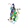
| Entry | Database: PDB / ID: 2prt | ||||||
|---|---|---|---|---|---|---|---|
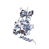
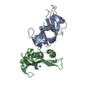
| Title | Structure of the Wilms Tumor Suppressor Protein Zinc Finger Domain Bound to DNA | ||||||
 Components Components |
| ||||||
 Keywords Keywords | TRANSCRIPTION/DNA / Protein-DNA complex / TRANSCRIPTION-DNA COMPLEX | ||||||
| Function / homology |  Function and homology information Function and homology informationposterior mesonephric tubule development / negative regulation of metanephric glomerular mesangial cell proliferation / positive regulation of metanephric ureteric bud development / thorax and anterior abdomen determination / metanephric epithelium development / regulation of animal organ formation / adrenal cortex formation / negative regulation of female gonad development / positive regulation of heart growth / visceral serous pericardium development ...posterior mesonephric tubule development / negative regulation of metanephric glomerular mesangial cell proliferation / positive regulation of metanephric ureteric bud development / thorax and anterior abdomen determination / metanephric epithelium development / regulation of animal organ formation / adrenal cortex formation / negative regulation of female gonad development / positive regulation of heart growth / visceral serous pericardium development / Nephron development / glomerular basement membrane development / diaphragm development / sex determination / positive regulation of male gonad development / cellular response to gonadotropin stimulus / Transcriptional regulation of testis differentiation / gonad development / metanephric mesenchyme development / metanephric S-shaped body morphogenesis / podocyte differentiation / double-stranded methylated DNA binding / tissue development / cardiac muscle cell fate commitment / mesenchymal to epithelial transition / hemi-methylated DNA-binding / male genitalia development / glomerulus development / camera-type eye development / C2H2 zinc finger domain binding / ureteric bud development / adrenal gland development / branching involved in ureteric bud morphogenesis / negative regulation of gene expression via chromosomal CpG island methylation / germ cell development / vasculogenesis / epithelial cell differentiation / RNA splicing / cellular response to cAMP / kidney development / negative regulation of cell growth / Negative Regulation of CDH1 Gene Transcription / positive regulation of miRNA transcription / male gonad development / heart development / DNA-binding transcription activator activity, RNA polymerase II-specific / sequence-specific DNA binding / DNA-binding transcription factor activity, RNA polymerase II-specific / nuclear speck / negative regulation of translation / transcription cis-regulatory region binding / RNA polymerase II cis-regulatory region sequence-specific DNA binding / positive regulation of apoptotic process / DNA-binding transcription factor activity / negative regulation of cell population proliferation / negative regulation of DNA-templated transcription / positive regulation of gene expression / regulation of transcription by RNA polymerase II / regulation of DNA-templated transcription / negative regulation of apoptotic process / positive regulation of DNA-templated transcription / nucleolus / DNA-templated transcription / negative regulation of transcription by RNA polymerase II / positive regulation of transcription by RNA polymerase II / RNA binding / zinc ion binding / nucleoplasm / nucleus / cytoplasm / cytosol Similarity search - Function | ||||||
| Biological species |  Homo sapiens (human) Homo sapiens (human) | ||||||
| Method |  X-RAY DIFFRACTION / SYNCHROTRON / MOLECULAR REPLACEMENT / Resolution: 3.15 Å X-RAY DIFFRACTION / SYNCHROTRON / MOLECULAR REPLACEMENT / Resolution: 3.15 Å | ||||||
 Authors Authors | Stoll, R. / Lee, B.M. / Debler, E.W. / Laity, J.H. / Wilson, I.A. / Dyson, H.J. / Wright, P.E. | ||||||
 Citation Citation | Journal: J.Mol.Biol. / Year: 2007 Title: Structure of the Wilms tumor suppressor protein zinc finger domain bound to DNA Authors: Stoll, R. / Lee, B.M. / Debler, E.W. / Laity, J.H. / Wilson, I.A. / Dyson, H.J. / Wright, P.E. | ||||||
| History |
|
- Structure visualization
| Structure viewer | Molecule: MolmilJmol/JSmol |
|---|
- Downloads & links
-Download
| PDBx/mmCIF format | 2prt.cif.gz | 51.6 KB | Display | PDBx/mmCIF format |
|---|---|---|---|---|
| PDB format | pdb2prt.ent.gz | 33.8 KB | Display | PDB format |
| PDBx/mmJSON format | 2prt.json.gz | Tree view | PDBx/mmJSON format | |
| Others |  Other downloads Other downloads |
-Validation report
| Arichive directory | https://data.pdbj.org/pub/pdb/validation_reports/pr/2prtftp://data.pdbj.org/pub/pdb/validation_reports/pr/2prt | HTTPS FTP |
|---|
-Related structure data
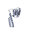
| Related structure data |  2jp9C  2jpaC  1aayS S: Starting model for refinement C: citing same article ( |
|---|---|
| Similar structure data |


-Links
 PDBj
PDBj


- Assembly
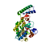
| Deposited unit | 
| ||||||||
|---|---|---|---|---|---|---|---|---|---|
| 1 |
| ||||||||
| Unit cell |
|
-Components
| #1: DNA chain | Mass: 4353.801 Da / Num. of mol.: 1 / Source method: obtained synthetically |
|---|---|
| #2: DNA chain | Mass: 4211.734 Da / Num. of mol.: 1 / Source method: obtained synthetically |
| #3: Protein | Mass: 14488.708 Da / Num. of mol.: 1 / Fragment: residues 174-291 Source method: isolated from a genetically manipulated source Source: (gene. exp.) Homo sapiens (human) / Gene: WT1 / Production host:  |
| #4: Chemical | ChemComp-ZN /   Mass: 65.409 Da / Num. of mol.: 4 / Source method: obtained synthetically / Formula: Zn Mass: 65.409 Da / Num. of mol.: 4 / Source method: obtained synthetically / Formula: Zn |
-Experimental details
-Experiment
| Experiment | Method: X-RAY DIFFRACTION / Number of used crystals: 1 |
|---|
- Sample preparation
| Crystal | Density Matthews: 3.97 Å3/Da / Density % sol: 69.04 % | ||||||||||||||||||||||||||||||||||||
|---|---|---|---|---|---|---|---|---|---|---|---|---|---|---|---|---|---|---|---|---|---|---|---|---|---|---|---|---|---|---|---|---|---|---|---|---|---|
| Crystal grow | Temperature: 277 K / pH: 6.7 Details: 10 mM d11-Tris/HCl, 20 mM KCl, 5uM ZnSO4, 2mM NaN3, 10% D2O, pH 6.7, SMALL TUBES, temperature 277K, pH 6.70 | ||||||||||||||||||||||||||||||||||||
| Components of the solutions |
|
-Data collection
| Diffraction | Mean temperature: 97 K |
|---|---|
| Diffraction source | Source: SYNCHROTRON / Site: SSRL  / Beamline: BL9-2 / Wavelength: 1.1921 / Beamline: BL9-2 / Wavelength: 1.1921 |
| Detector | Date: Feb 1, 2002 |
| Radiation | Protocol: SINGLE WAVELENGTH / Monochromatic (M) / Laue (L): M / Scattering type: x-ray |
| Radiation wavelength | Wavelength: 1.1921 Å / Relative weight: 1 |
| Reflection | Resolution: 3.15→42.8 Å / Num. obs: 7166 / % possible obs: 99.1 % / Redundancy: 11.2 % / Rsym value: 0.085 / Net I/σ(I): 22.9 |
| Reflection shell | Highest resolution: 3.15 Å / Redundancy: 11.7 % / Mean I/σ(I) obs: 5.3 / Rsym value: 0.435 / % possible all: 100 |
- Processing
| Software | Name: CNS / Classification: refinement | ||||||||||||
|---|---|---|---|---|---|---|---|---|---|---|---|---|---|
| Refinement | Method to determine structure: MOLECULAR REPLACEMENT Starting model: PDB entry 1AAY Highest resolution: 3.15 Å
| ||||||||||||
| Refinement step | Cycle: LAST / Highest resolution: 3.15 Å
| ||||||||||||
| LS refinement shell | Highest resolution: 3.15 Å / Rfactor Rfree: 0.405 / Rfactor Rwork: 0.376 |
