 Movie
Movie Controller

[English] 日本語
 Yorodumi
- PDB-2v9l: L-RHAMNULOSE-1-PHOSPHATE ALDOLASE FROM ESCHERICHIA COLI (MUTANT Q... -
+ Open data

- Basic information
| Entry | Database: PDB / ID: 2v9l | ||||||
|---|---|---|---|---|---|---|---|
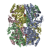
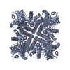
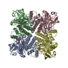
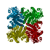
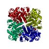
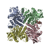
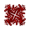
| Title | L-RHAMNULOSE-1-PHOSPHATE ALDOLASE FROM ESCHERICHIA COLI (MUTANT Q6Y- E192A) | ||||||
 Components Components | RHAMNULOSE-1-PHOSPHATE ALDOLASE | ||||||
 Keywords Keywords | LYASE / ENTROPY INDEX / METAL-BINDING / OLIGOMERIZATION / ZINC / ALDOLASE / CLASS II / CYTOPLASM / CLEAVAGE OF L-RHAMNULOSE-1-PHOSPHATE TO DIHYDROXYACETONEPH BACTERIAL L-RHAMNOSE METABOLISM / INTERFACE DESIGN / SURFACE MUTATION / 2-KETOSE DEGRADATION / PROTEIN-PROTEIN INTERFACE / RARE SUGAR / AGGREGATION / ZINC ENZYME / FIBRILLATION / RHAMNOSE METABOLISM / PROTEIN ENGINEERING | ||||||
| Function / homology |  Function and homology information Function and homology informationrhamnulose-1-phosphate aldolase / rhamnulose-1-phosphate aldolase activity / rhamnose catabolic process / pentose catabolic process / aldehyde-lyase activity / metal ion binding / identical protein binding / cytosol Similarity search - Function | ||||||
| Biological species |  | ||||||
| Method |  X-RAY DIFFRACTION / SYNCHROTRON / MOLECULAR REPLACEMENT / Resolution: 1.23 Å X-RAY DIFFRACTION / SYNCHROTRON / MOLECULAR REPLACEMENT / Resolution: 1.23 Å | ||||||
 Authors Authors | Grueninger, D. / Schulz, G.E. | ||||||
 Citation Citation | Journal: Science / Year: 2008 Title: Designed Protein-Protein Association. Authors: Grueninger, D. / Treiber, N. / Ziegler, M.O.P. / Koetter, J.W.A. / Schulze, M.-S. / Schulz, G.E. #1: Journal: Biochemistry / Year: 2003Title: Structure and Catalytic Mechanism of L-Rhamnulose-1-Phosphate Aldolase Authors: Kroemer, M. / Merkel, I. / Schulz, G.E. | ||||||
| History |
|
- Structure visualization
| Structure viewer | Molecule: MolmilJmol/JSmol |
|---|
- Downloads & links
-Download
| PDBx/mmCIF format | 2v9l.cif.gz | 208.4 KB | Display | PDBx/mmCIF format |
|---|---|---|---|---|
| PDB format | pdb2v9l.ent.gz | 170.3 KB | Display | PDB format |
| PDBx/mmJSON format | 2v9l.json.gz | Tree view | PDBx/mmJSON format | |
| Others |  Other downloads Other downloads |
-Validation report
| Arichive directory | https://data.pdbj.org/pub/pdb/validation_reports/v9/2v9lftp://data.pdbj.org/pub/pdb/validation_reports/v9/2v9l | HTTPS FTP |
|---|
-Related structure data
| Related structure data |  2uyuC  2uyvC  2v7gC  2v9eC  2v9fC  2v9gC  2v9iC  2v9mC  2v9nC  2v9oC  2v9uC  1ojrS S: Starting model for refinement C: citing same article ( |
|---|---|
| Similar structure data |


-Links
 PDBj
PDBj- Assembly
| Deposited unit | 
| ||||||||||||||||||||||||
|---|---|---|---|---|---|---|---|---|---|---|---|---|---|---|---|---|---|---|---|---|---|---|---|---|---|
| 1 | 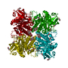
| ||||||||||||||||||||||||
| Unit cell |
| ||||||||||||||||||||||||
| Components on special symmetry positions |
|
-Components
-Protein , 1 types, 1 molecules A
| #1: Protein | Mass: 30151.416 Da / Num. of mol.: 1 / Mutation: YES Source method: isolated from a genetically manipulated source Source: (gene. exp.) References: UniProt: P32169, rhamnulose-1-phosphate aldolase |
|---|
-Non-polymers , 5 types, 578 molecules 


| #2: Chemical | ChemComp-ZN /  Mass: 65.409 Da / Num. of mol.: 1 / Source method: obtained synthetically / Formula: Zn Mass: 65.409 Da / Num. of mol.: 1 / Source method: obtained synthetically / Formula: Zn | ||||
|---|---|---|---|---|---|
| #3: Chemical | ChemComp-PO4 /  Mass: 94.971 Da / Num. of mol.: 1 / Source method: obtained synthetically / Formula: PO4 Mass: 94.971 Da / Num. of mol.: 1 / Source method: obtained synthetically / Formula: PO4 | ||||
| #4: Chemical | ChemComp-ACT /  Mass: 59.044 Da / Num. of mol.: 4 / Source method: obtained synthetically / Formula: C2H3O2 Mass: 59.044 Da / Num. of mol.: 4 / Source method: obtained synthetically / Formula: C2H3O2#5: Chemical | ChemComp-PGO /  Mass: 76.094 Da / Num. of mol.: 8 / Source method: obtained synthetically / Formula: C3H8O2 Mass: 76.094 Da / Num. of mol.: 8 / Source method: obtained synthetically / Formula: C3H8O2#6: Water | ChemComp-HOH / | Mass: 18.015 Da / Num. of mol.: 564 / Source method: isolated from a natural source / Formula: H2O |
-Details
| Compound details | ENGINEERED |
|---|
-Experimental details
-Experiment
| Experiment | Method: X-RAY DIFFRACTION / Number of used crystals: 1 |
|---|
- Sample preparation
| Crystal | Density Matthews: 2.7 Å3/Da / Density % sol: 54.8 % / Description: NONE |
|---|---|
| Crystal grow | pH: 5 Details: 40% (V/V) 1,2-PROPANEDIOL, 0.1 M ACETATE (PH4.5), pH 5.0 |
-Data collection
| Diffraction | Mean temperature: 100 K |
|---|---|
| Diffraction source | Source: SYNCHROTRON / Site: EMBL/DESY, HAMBURG  / Beamline: X11 / Wavelength: 0.8123 / Beamline: X11 / Wavelength: 0.8123 |
| Detector | Type: MARRESEARCH / Detector: CCD / Date: Oct 14, 2003 |
| Radiation | Protocol: SINGLE WAVELENGTH / Monochromatic (M) / Laue (L): M / Scattering type: x-ray |
| Radiation wavelength | Wavelength: 0.8123 Å / Relative weight: 1 |
| Reflection | Resolution: 1.23→20 Å / Num. obs: 96326 / % possible obs: 99.4 % / Redundancy: 8.81 % / Rmerge(I) obs: 0.06 / Net I/σ(I): 20 |
| Reflection shell | Highest resolution: 1.23 Å / Redundancy: 8.83 % / Rmerge(I) obs: 0.36 / Mean I/σ(I) obs: 6.15 / % possible all: 100 |
- Processing
| Software |
| |||||||||||||||||||||||||||||||||
|---|---|---|---|---|---|---|---|---|---|---|---|---|---|---|---|---|---|---|---|---|---|---|---|---|---|---|---|---|---|---|---|---|---|---|
| Refinement | Method to determine structure: MOLECULAR REPLACEMENT Starting model: 1OJR Resolution: 1.23→10 Å / Num. parameters: 26231 / Num. restraintsaints: 32549 / Cross valid method: FREE R-VALUE / σ(F): 0 / Stereochemistry target values: ENGH AND HUBER
| |||||||||||||||||||||||||||||||||
| Refine analyze | Num. disordered residues: 39 / Occupancy sum hydrogen: 2152.5 / Occupancy sum non hydrogen: 2712.73 | |||||||||||||||||||||||||||||||||
| Refinement step | Cycle: LAST / Resolution: 1.23→10 Å
| |||||||||||||||||||||||||||||||||
| Refine LS restraints |
|
