 Movie
Movie Controller

[English] 日本語
 Yorodumi
- PDB-2uyu: L-RHAMNULOSE-1-PHOSPHATE ALDOLASE FROM ESCHERICHIA COLI (MUTANT A... -
+ Open data

- Basic information
| Entry | Database: PDB / ID: 2uyu | ||||||
|---|---|---|---|---|---|---|---|
| Title | L-RHAMNULOSE-1-PHOSPHATE ALDOLASE FROM ESCHERICHIA COLI (MUTANT A88F- E192A) | ||||||
 Components Components | RHAMNULOSE-1-PHOSPHATE ALDOLASE | ||||||
 Keywords Keywords | LYASE / AGGREGATION / ZINC ENZYME / FIBRILLATION / METAL-BINDING / OLIGOMERIZATION / INTERFACE DESIGN / SURFACE MUTATION / 2-KETOSE DEGRADATION / PROTEIN-PROTEIN INTERFACE / ZINC / ALDOLASE / CLASS II / RARE SUGAR / CLEAVAGE OF L-RHAMNULOSE-1-PHOSPHATE TO DIHYDROXYACETONEPH BACTERIAL L-RHAMNOSE METABOLISM / RHAMNOSE METABOLISM / PROTEIN ENGINEERING | ||||||
| Function / homology |  Function and homology information Function and homology informationrhamnulose-1-phosphate aldolase / rhamnulose-1-phosphate aldolase activity / rhamnose catabolic process / pentose catabolic process / aldehyde-lyase activity / metal ion binding / identical protein binding / cytosol Similarity search - Function | ||||||
| Biological species |  | ||||||
| Method |  X-RAY DIFFRACTION / SYNCHROTRON / MOLECULAR REPLACEMENT / Resolution: 1.96 Å X-RAY DIFFRACTION / SYNCHROTRON / MOLECULAR REPLACEMENT / Resolution: 1.96 Å | ||||||
 Authors Authors | Grueninger, D. / Schulz, G.E. | ||||||
 Citation Citation | Journal: Science / Year: 2008 Title: Designed Protein-Protein Association. Authors: Grueninger, D. / Treiber, N. / Ziegler, M.O.P. / Koetter, J.W.A. / Schulze, M.-S. / Schulz, G.E. #1: Journal: Biochemistry / Year: 2003Title: Structure and Catalytic Mechanism of L-Rhamnulose-1-Phosphate Aldolase Authors: Kroemer, M. / Merkel, I. / Schulz, G.E. | ||||||
| History |
|
- Structure visualization
| Structure viewer | Molecule: MolmilJmol/JSmol |
|---|
- Downloads & links
-Download
| PDBx/mmCIF format | 2uyu.cif.gz | 127.6 KB | Display | PDBx/mmCIF format |
|---|---|---|---|---|
| PDB format | pdb2uyu.ent.gz | 99.5 KB | Display | PDB format |
| PDBx/mmJSON format | 2uyu.json.gz | Tree view | PDBx/mmJSON format | |
| Others |  Other downloads Other downloads |
-Validation report
| Arichive directory | https://data.pdbj.org/pub/pdb/validation_reports/uy/2uyuftp://data.pdbj.org/pub/pdb/validation_reports/uy/2uyu | HTTPS FTP |
|---|
-Related structure data
| Related structure data |  2uyvC  2v7gC  2v9eC  2v9fC  2v9gC  2v9iC  2v9lC  2v9mC  2v9nC  2v9oC  2v9uC  1ojrS S: Starting model for refinement C: citing same article ( |
|---|---|
| Similar structure data |


-Links
 PDBj
PDBj- Assembly
| Deposited unit | 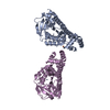
| ||||||||||||||||||
|---|---|---|---|---|---|---|---|---|---|---|---|---|---|---|---|---|---|---|---|
| 1 | x 8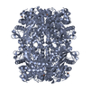
| ||||||||||||||||||
| 2 | x 8
| ||||||||||||||||||
| Unit cell |
| ||||||||||||||||||
| Components on special symmetry positions |
| ||||||||||||||||||
| Noncrystallographic symmetry (NCS) | NCS domain:
NCS domain segments: Component-ID: 1 / Ens-ID: 1 / Beg auth comp-ID: MET / Beg label comp-ID: MET / End auth comp-ID: LEU / End label comp-ID: LEU / Refine code: 1 / Auth seq-ID: 1 - 274 / Label seq-ID: 1 - 274
NCS oper: (Code: given Matrix: (-0.7072, -0.707, 0.000623), Vector: |
-Components
| #1: Protein | Mass: 30192.467 Da / Num. of mol.: 2 / Mutation: YES Source method: isolated from a genetically manipulated source Source: (gene. exp.) References: UniProt: P32169, rhamnulose-1-phosphate aldolase #2: Chemical |   Mass: 65.409 Da / Num. of mol.: 2 / Source method: obtained synthetically / Formula: Zn Mass: 65.409 Da / Num. of mol.: 2 / Source method: obtained synthetically / Formula: Zn#3: Chemical |   Mass: 55.845 Da / Num. of mol.: 2 / Source method: obtained synthetically / Formula: Fe Mass: 55.845 Da / Num. of mol.: 2 / Source method: obtained synthetically / Formula: Fe#4: Water | ChemComp-HOH / |  Mass: 18.015 Da / Num. of mol.: 341 / Source method: isolated from a natural source / Formula: H2O Mass: 18.015 Da / Num. of mol.: 341 / Source method: isolated from a natural source / Formula: H2OCompound details | ENGINEERED RESIDUE IN CHAIN A, ALA 88 TO PHE ENGINEERED RESIDUE IN CHAIN A, GLU 192 TO ALA ...ENGINEERED | |
|---|
-Experimental details
-Experiment
| Experiment | Method: X-RAY DIFFRACTION |
|---|
- Sample preparation
| Crystal | Density Matthews: 2.7 Å3/Da / Density % sol: 54.1 % / Description: NONE |
|---|---|
| Crystal grow | pH: 7.4 Details: 40% (V/V) ETHYLENE GLYCOL, 100 MM HEPES (PH 7.5), 5% (W/V) PEG 3000, RESULTING PH: 7.4 |
-Data collection
| Diffraction | Mean temperature: 100 K |
|---|---|
| Diffraction source | Source: SYNCHROTRON / Site: EMBL/DESY, HAMBURG  / Beamline: X11 / Wavelength: 0.8123 / Beamline: X11 / Wavelength: 0.8123 |
| Detector | Type: MARRESEARCH / Detector: CCD / Date: Oct 14, 2003 |
| Radiation | Protocol: SINGLE WAVELENGTH / Monochromatic (M) / Laue (L): M / Scattering type: x-ray |
| Radiation wavelength | Wavelength: 0.8123 Å / Relative weight: 1 |
| Reflection | Resolution: 1.96→20 Å / Num. obs: 47844 / % possible obs: 99.7 % / Observed criterion σ(I): 3.9 / Redundancy: 4.84 % / Rmerge(I) obs: 0.06 / Net I/σ(I): 16.47 |
| Reflection shell | Resolution: 1.96→1.99 Å / Redundancy: 4.89 % / Rmerge(I) obs: 0.41 / Mean I/σ(I) obs: 3.9 / % possible all: 100 |
- Processing
| Software |
| ||||||||||||||||||||||||||||||||||||||||||||||||||||||||||||||||||||||||||||||||||||||||||||||||||||||||||||||||||||||||||||||||||||||||||||||||||||||||||||||||||||||||||||||||||||||
|---|---|---|---|---|---|---|---|---|---|---|---|---|---|---|---|---|---|---|---|---|---|---|---|---|---|---|---|---|---|---|---|---|---|---|---|---|---|---|---|---|---|---|---|---|---|---|---|---|---|---|---|---|---|---|---|---|---|---|---|---|---|---|---|---|---|---|---|---|---|---|---|---|---|---|---|---|---|---|---|---|---|---|---|---|---|---|---|---|---|---|---|---|---|---|---|---|---|---|---|---|---|---|---|---|---|---|---|---|---|---|---|---|---|---|---|---|---|---|---|---|---|---|---|---|---|---|---|---|---|---|---|---|---|---|---|---|---|---|---|---|---|---|---|---|---|---|---|---|---|---|---|---|---|---|---|---|---|---|---|---|---|---|---|---|---|---|---|---|---|---|---|---|---|---|---|---|---|---|---|---|---|---|---|
| Refinement | Method to determine structure: MOLECULAR REPLACEMENT Starting model: PDB ENTRY 1OJR Resolution: 1.96→19.96 Å / Cor.coef. Fo:Fc: 0.949 / Cor.coef. Fo:Fc free: 0.923 / SU B: 8.479 / SU ML: 0.129 / Cross valid method: THROUGHOUT / ESU R: 0.171 / ESU R Free: 0.159 / Stereochemistry target values: MAXIMUM LIKELIHOOD / Details: HYDROGENS HAVE BEEN ADDED IN THE RIDING POSITIONS.
| ||||||||||||||||||||||||||||||||||||||||||||||||||||||||||||||||||||||||||||||||||||||||||||||||||||||||||||||||||||||||||||||||||||||||||||||||||||||||||||||||||||||||||||||||||||||
| Solvent computation | Ion probe radii: 0.8 Å / Shrinkage radii: 0.8 Å / VDW probe radii: 1.4 Å / Solvent model: MASK | ||||||||||||||||||||||||||||||||||||||||||||||||||||||||||||||||||||||||||||||||||||||||||||||||||||||||||||||||||||||||||||||||||||||||||||||||||||||||||||||||||||||||||||||||||||||
| Displacement parameters | Biso mean: 27.21 Å2
| ||||||||||||||||||||||||||||||||||||||||||||||||||||||||||||||||||||||||||||||||||||||||||||||||||||||||||||||||||||||||||||||||||||||||||||||||||||||||||||||||||||||||||||||||||||||
| Refinement step | Cycle: LAST / Resolution: 1.96→19.96 Å
| ||||||||||||||||||||||||||||||||||||||||||||||||||||||||||||||||||||||||||||||||||||||||||||||||||||||||||||||||||||||||||||||||||||||||||||||||||||||||||||||||||||||||||||||||||||||
| Refine LS restraints |
|