 Movie
Movie Controller

[English] 日本語
 Yorodumi
- PDB-2v8y: Crystallographic and mass spectrometric characterisation of eIF4E... -
+ Open data

- Basic information
| Entry | Database: PDB / ID: 2v8y | ||||||
|---|---|---|---|---|---|---|---|
| Title | Crystallographic and mass spectrometric characterisation of eIF4E with N7-cap derivatives | ||||||
 Components Components |
| ||||||
 Keywords Keywords | TRANSLATION / PHOSPHORYLATION / INITIATION FACTOR / CAP / EIF4E / 7BNGMP / 4E-BP1 / RNA-BINDING / ACETYLATION / HOST-VIRUS INTERACTION / PROTEIN SYNTHESIS INHIBITOR / PROTEIN BIOSYNTHESIS / TRANSLATION REGULATION | ||||||
| Function / homology |  Function and homology information Function and homology informationeukaryotic initiation factor 4G binding / Activation of the mRNA upon binding of the cap-binding complex and eIFs, and subsequent binding to 43S / eukaryotic initiation factor 4E binding / regulation of translation at postsynapse, modulating synaptic transmission / RNA cap binding / eukaryotic translation initiation factor 4F complex / Z-decay: degradation of maternal mRNAs by zygotically expressed factors / chromatoid body / Deadenylation of mRNA / mRNA cap binding ...eukaryotic initiation factor 4G binding / Activation of the mRNA upon binding of the cap-binding complex and eIFs, and subsequent binding to 43S / eukaryotic initiation factor 4E binding / regulation of translation at postsynapse, modulating synaptic transmission / RNA cap binding / eukaryotic translation initiation factor 4F complex / Z-decay: degradation of maternal mRNAs by zygotically expressed factors / chromatoid body / Deadenylation of mRNA / mRNA cap binding / Transport of the SLBP independent Mature mRNA / Transport of the SLBP Dependant Mature mRNA / RNA 7-methylguanosine cap binding / Transport of Mature mRNA Derived from an Intronless Transcript / M-decay: degradation of maternal mRNAs by maternally stored factors / RISC complex / negative regulation of neuron differentiation / stem cell population maintenance / Ribosomal scanning and start codon recognition / Translation initiation complex formation / TOR signaling / mTORC1-mediated signalling / cellular response to dexamethasone stimulus / behavioral fear response / GTP hydrolysis and joining of the 60S ribosomal subunit / mRNA export from nucleus / L13a-mediated translational silencing of Ceruloplasmin expression / positive regulation of mitotic cell cycle / translation initiation factor activity / translation repressor activity / negative regulation of translational initiation / translation initiation factor binding / G1/S transition of mitotic cell cycle / translational initiation / P-body / ISG15 antiviral mechanism / neuron differentiation / cytoplasmic stress granule / cytoplasmic ribonucleoprotein granule / regulation of translation / DNA-binding transcription factor binding / postsynapse / nuclear speck / negative regulation of translation / perinuclear region of cytoplasm / glutamatergic synapse / enzyme binding / RNA binding / extracellular exosome / nucleoplasm / nucleus / cytosol / cytoplasm Similarity search - Function | ||||||
| Biological species |  HOMO SAPIENS (human) HOMO SAPIENS (human) | ||||||
| Method |  X-RAY DIFFRACTION / SYNCHROTRON / MOLECULAR REPLACEMENT / Resolution: 2.1 Å X-RAY DIFFRACTION / SYNCHROTRON / MOLECULAR REPLACEMENT / Resolution: 2.1 Å | ||||||
 Authors Authors | Brown, C.J. / Mcnae, I. / Fischer, P.M. / Walkinshaw, M.D. | ||||||
 Citation Citation | Journal: J.Mol.Biol. / Year: 2007 Title: Crystallographic and Mass Spectrometric Characterisation of Eif4E with N(7)-Alkylated CAP Derivatives. Authors: Brown, C.J. / Mcnae, I. / Fischer, P.M. / Walkinshaw, M.D. | ||||||
| History |
|
- Structure visualization
| Structure viewer | Molecule: MolmilJmol/JSmol |
|---|
- Downloads & links
-Download
| PDBx/mmCIF format | 2v8y.cif.gz | 103.1 KB | Display | PDBx/mmCIF format |
|---|---|---|---|---|
| PDB format | pdb2v8y.ent.gz | 78.5 KB | Display | PDB format |
| PDBx/mmJSON format | 2v8y.json.gz | Tree view | PDBx/mmJSON format | |
| Others |  Other downloads Other downloads |
-Validation report
| Arichive directory | https://data.pdbj.org/pub/pdb/validation_reports/v8/2v8yftp://data.pdbj.org/pub/pdb/validation_reports/v8/2v8y | HTTPS FTP |
|---|
-Related structure data
| Related structure data |  2v8wC  2v8xC  1ejhS C: citing same article ( S: Starting model for refinement |
|---|---|
| Similar structure data |


-Links
 PDBj
PDBj


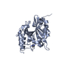
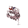
- Assembly
| Deposited unit | 
| ||||||||||||
|---|---|---|---|---|---|---|---|---|---|---|---|---|---|
| 1 | 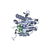
| ||||||||||||
| 2 | 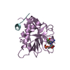
| ||||||||||||
| Unit cell |
| ||||||||||||
| Noncrystallographic symmetry (NCS) | NCS oper:
|
-Components
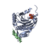
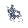
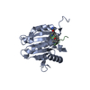
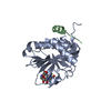
| #1: Protein | Mass: 25130.242 Da / Num. of mol.: 2 Source method: isolated from a genetically manipulated source Source: (gene. exp.) HOMO SAPIENS (human) / Plasmid: PET11D / Production host:  #2: Protein/peptide | Mass: 1861.240 Da / Num. of mol.: 2 / Fragment: PHAS-I4E-BP1 BINDING PEPTIDE, RESIDUES 50-63 / Source method: obtained synthetically / Source: (synth.) HOMO SAPIENS (human) / References: UniProt: Q13541#3: Chemical | 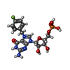  Type: RNA linking / Mass: 472.342 Da / Num. of mol.: 2 / Source method: obtained synthetically / Formula: C17H20FN5O8P Type: RNA linking / Mass: 472.342 Da / Num. of mol.: 2 / Source method: obtained synthetically / Formula: C17H20FN5O8P#4: Water | ChemComp-HOH / |  Mass: 18.015 Da / Num. of mol.: 312 / Source method: isolated from a natural source / Formula: H2O Mass: 18.015 Da / Num. of mol.: 312 / Source method: isolated from a natural source / Formula: H2O |
|---|
-Experimental details
-Experiment
| Experiment | Method: X-RAY DIFFRACTION / Number of used crystals: 1 |
|---|
- Sample preparation
| Crystal | Density Matthews: 2.62 Å3/Da / Density % sol: 53.08 % / Description: NONE |
|---|---|
| Crystal grow | pH: 7 Details: 25% PEG 6000, 7% AMMONIUM SULPHATE, 100MM HEPES-KOH PH 7.0 |
-Data collection
| Diffraction | Mean temperature: 100 K |
|---|---|
| Diffraction source | Source: SYNCHROTRON / Site: SRS  / Beamline: PX14.1 / Wavelength: 1.48 / Beamline: PX14.1 / Wavelength: 1.48 |
| Detector | Type: ADSC CCD / Detector: CCD / Details: MIRRORS |
| Radiation | Monochromator: SI 111 / Protocol: SINGLE WAVELENGTH / Monochromatic (M) / Laue (L): M / Scattering type: x-ray |
| Radiation wavelength | Wavelength: 1.48 Å / Relative weight: 1 |
| Reflection | Resolution: 2.1→41.17 Å / Num. obs: 28510 / % possible obs: 91.1 % / Observed criterion σ(I): 2 / Redundancy: 3.9 % / Rmerge(I) obs: 0.12 / Net I/σ(I): 2 |
| Reflection shell | Resolution: 2.1→2.21 Å / Redundancy: 3.9 % / Rmerge(I) obs: 0.53 / Mean I/σ(I) obs: 2.3 / % possible all: 87.6 |
- Processing
| Software |
| ||||||||||||||||||||||||||||||||||||||||||||||||||||||||||||||||||||||||||||||||||||||||||||||||||||||||||||||||||||||||||||||||||||||||||||||||||||||||||||||||||||||||||||||||||||||
|---|---|---|---|---|---|---|---|---|---|---|---|---|---|---|---|---|---|---|---|---|---|---|---|---|---|---|---|---|---|---|---|---|---|---|---|---|---|---|---|---|---|---|---|---|---|---|---|---|---|---|---|---|---|---|---|---|---|---|---|---|---|---|---|---|---|---|---|---|---|---|---|---|---|---|---|---|---|---|---|---|---|---|---|---|---|---|---|---|---|---|---|---|---|---|---|---|---|---|---|---|---|---|---|---|---|---|---|---|---|---|---|---|---|---|---|---|---|---|---|---|---|---|---|---|---|---|---|---|---|---|---|---|---|---|---|---|---|---|---|---|---|---|---|---|---|---|---|---|---|---|---|---|---|---|---|---|---|---|---|---|---|---|---|---|---|---|---|---|---|---|---|---|---|---|---|---|---|---|---|---|---|---|---|
| Refinement | Method to determine structure: MOLECULAR REPLACEMENT Starting model: PDB ENTRY 1EJH Resolution: 2.1→25.3 Å / Cor.coef. Fo:Fc: 0.935 / Cor.coef. Fo:Fc free: 0.903 / SU B: 4.956 / SU ML: 0.135 / Cross valid method: THROUGHOUT / ESU R: 0.242 / ESU R Free: 0.203 / Stereochemistry target values: MAXIMUM LIKELIHOOD / Details: HYDROGENS HAVE BEEN ADDED IN THE RIDING POSITIONS.
| ||||||||||||||||||||||||||||||||||||||||||||||||||||||||||||||||||||||||||||||||||||||||||||||||||||||||||||||||||||||||||||||||||||||||||||||||||||||||||||||||||||||||||||||||||||||
| Solvent computation | Ion probe radii: 0.8 Å / Shrinkage radii: 0.8 Å / VDW probe radii: 1.4 Å / Solvent model: MASK | ||||||||||||||||||||||||||||||||||||||||||||||||||||||||||||||||||||||||||||||||||||||||||||||||||||||||||||||||||||||||||||||||||||||||||||||||||||||||||||||||||||||||||||||||||||||
| Displacement parameters | Biso mean: 24.21 Å2
| ||||||||||||||||||||||||||||||||||||||||||||||||||||||||||||||||||||||||||||||||||||||||||||||||||||||||||||||||||||||||||||||||||||||||||||||||||||||||||||||||||||||||||||||||||||||
| Refinement step | Cycle: LAST / Resolution: 2.1→25.3 Å
| ||||||||||||||||||||||||||||||||||||||||||||||||||||||||||||||||||||||||||||||||||||||||||||||||||||||||||||||||||||||||||||||||||||||||||||||||||||||||||||||||||||||||||||||||||||||
| Refine LS restraints |
|