[English] 日本語
Yorodumi
- PDB-4bea: Crystal Structure of eIF4E in Complex with a Stapled Peptide Deri... -

+
Open data


ID or keywords:

Loading...

-
Basic information

Entry
Database: PDB / ID: 4bea
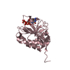
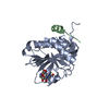
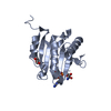
TitleCrystal Structure of eIF4E in Complex with a Stapled Peptide Derivative
Components
  • Eukaryotic translation initiation factor 4E
  • STAPLED EIF4E INTERACTING PEPTIDE
KeywordsTRANSLATION
Function / homology
Function and homology information


Activation of the mRNA upon binding of the cap-binding complex and eIFs, and subsequent binding to 43S / eukaryotic initiation factor 4G binding / regulation of translation at postsynapse, modulating synaptic transmission / RNA cap binding / chromatoid body / eukaryotic translation initiation factor 4F complex / Z-decay: degradation of maternal mRNAs by zygotically expressed factors / mRNA cap binding / Deadenylation of mRNA / Transport of the SLBP independent Mature mRNA ...Activation of the mRNA upon binding of the cap-binding complex and eIFs, and subsequent binding to 43S / eukaryotic initiation factor 4G binding / regulation of translation at postsynapse, modulating synaptic transmission / RNA cap binding / chromatoid body / eukaryotic translation initiation factor 4F complex / Z-decay: degradation of maternal mRNAs by zygotically expressed factors / mRNA cap binding / Deadenylation of mRNA / Transport of the SLBP independent Mature mRNA / Transport of the SLBP Dependant Mature mRNA / RNA 7-methylguanosine cap binding / M-decay: degradation of maternal mRNAs by maternally stored factors / Transport of Mature mRNA Derived from an Intronless Transcript / RISC complex / Ribosomal scanning and start codon recognition / stem cell population maintenance / Translation initiation complex formation / mTORC1-mediated signalling / negative regulation of neuron differentiation / GTP hydrolysis and joining of the 60S ribosomal subunit / L13a-mediated translational silencing of Ceruloplasmin expression / behavioral fear response / mRNA export from nucleus / translation initiation factor activity / positive regulation of mitotic cell cycle / cellular response to dexamethasone stimulus / translational initiation / P-body / neuron differentiation / ISG15 antiviral mechanism / cytoplasmic stress granule / cytoplasmic ribonucleoprotein granule / G1/S transition of mitotic cell cycle / regulation of translation / DNA-binding transcription factor binding / postsynapse / negative regulation of translation / nuclear speck / glutamatergic synapse / perinuclear region of cytoplasm / enzyme binding / RNA binding / extracellular exosome / nucleus / cytosol / cytoplasm
Similarity search - Function
RNA Cap, Translation Initiation Factor Eif4e / RNA Cap, Translation Initiation Factor Eif4e / Eukaryotic translation initiation factor 4E (eIF-4E), conserved site / Eukaryotic initiation factor 4E signature. / Translation Initiation factor eIF- 4e / Eukaryotic initiation factor 4E / Translation Initiation factor eIF- 4e-like / 2-Layer Sandwich / Alpha Beta
Similarity search - Domain/homology
Eukaryotic translation initiation factor 4E
Similarity search - Component
Biological speciesHomo sapiens (human)
SYNTHETIC CONSTRUCT (others)
MethodX-RAY DIFFRACTION / MOLECULAR REPLACEMENT / Resolution: 2.57 Å
AuthorsQuah, S.T. / Lama, D. / Verma, C.S. / Lane, D.P. / Brown, C.J.
CitationJournal: Sci.Rep. / Year: 2013
Title: Rational Optimization of Conformational Effects Induced by Hydrocarbon Staples in Peptides and Their Binding Interfaces.
Authors: Lama, D. / Quah, S.T. / Verma, C.S. / Lakshminarayanan, R. / Beuerman, R.W. / Lane, D.P. / Brown, C.J.
History
DepositionMar 7, 2013Deposition site: PDBE / Processing site: PDBE
Revision 1.0Dec 11, 2013Provider: repository / Type: Initial release
Revision 1.1Jan 8, 2014Group: Database references
Revision 1.2Apr 4, 2018Group: Data collection / Category: diffrn_source / Item: _diffrn_source.type
Revision 2.0Mar 15, 2023Group: Atomic model / Database references ...Atomic model / Database references / Derived calculations / Non-polymer description / Other / Polymer sequence / Source and taxonomy / Structure summary
Category: atom_site / chem_comp ...atom_site / chem_comp / database_2 / entity / entity_name_com / entity_poly / entity_poly_seq / entity_src_gen / pdbx_database_status / pdbx_entity_src_syn / pdbx_poly_seq_scheme / struct_conn / struct_ref
Item: _atom_site.auth_comp_id / _atom_site.label_comp_id ..._atom_site.auth_comp_id / _atom_site.label_comp_id / _chem_comp.formula / _chem_comp.formula_weight / _chem_comp.id / _chem_comp.name / _database_2.pdbx_DOI / _database_2.pdbx_database_accession / _entity.formula_weight / _entity.pdbx_description / _entity_name_com.name / _entity_poly.pdbx_seq_one_letter_code / _entity_poly_seq.mon_id / _entity_src_gen.gene_src_common_name / _entity_src_gen.pdbx_beg_seq_num / _entity_src_gen.pdbx_end_seq_num / _entity_src_gen.pdbx_gene_src_gene / _entity_src_gen.pdbx_gene_src_scientific_name / _entity_src_gen.pdbx_seq_type / _pdbx_database_status.status_code_sf / _pdbx_entity_src_syn.pdbx_beg_seq_num / _pdbx_entity_src_syn.pdbx_end_seq_num / _pdbx_poly_seq_scheme.mon_id / _pdbx_poly_seq_scheme.pdb_mon_id / _struct_conn.pdbx_dist_value / _struct_conn.pdbx_leaving_atom_flag / _struct_conn.ptnr1_auth_comp_id / _struct_conn.ptnr1_auth_seq_id / _struct_conn.ptnr1_label_atom_id / _struct_conn.ptnr1_label_comp_id / _struct_conn.ptnr1_label_seq_id / _struct_conn.ptnr2_auth_comp_id / _struct_conn.ptnr2_auth_seq_id / _struct_conn.ptnr2_label_atom_id / _struct_conn.ptnr2_label_comp_id / _struct_conn.ptnr2_label_seq_id / _struct_ref.pdbx_align_begin / _struct_ref.pdbx_seq_one_letter_code
Revision 2.1Feb 7, 2024Group: Data collection / Refinement description
Category: chem_comp_atom / chem_comp_bond / pdbx_initial_refinement_model

-
Structure visualization

Structure viewerMolecule:
MolmilJmol/JSmol

Downloads & links

-
Assembly

Deposited unit
A: Eukaryotic translation initiation factor 4E
B: STAPLED EIF4E INTERACTING PEPTIDE


Theoretical massNumber of molelcules
Total (without water)26,6912
Polymers26,6912
Non-polymers00
Water50428
1


  • Idetical with deposited unit
  • defined by author&software
TypeNameSymmetry operationNumber
identity operation1_555x,y,z1
Buried area1320 Å2
ΔGint-6.4 kcal/mol
Surface area10620 Å2
MethodPISA
Unit cell
Length a, b, c (Å)38.301, 91.677, 137.908
Angle α, β, γ (deg.)90.00, 90.00, 90.00
Int Tables number20
Space group name H-MC2221
Components on special symmetry positions
IDModelComponents
11B-2001-

HOH

-
Components

#1: Protein Eukaryotic translation initiation factor 4E / eIF4E / eIF-4F 25 kDa subunit / mRNA cap-binding protein


Mass: 25130.242 Da / Num. of mol.: 1
Source method: isolated from a genetically manipulated source
Source: (gene. exp.) Homo sapiens (human) / Gene: EIF4E, EIF4EL1, EIF4F / Production host: ESCHERICHIA COLI (E. coli) / Strain (production host): BL21(DE3) / References: UniProt: P06730
#2: Protein/peptide STAPLED EIF4E INTERACTING PEPTIDE


Mass: 1560.992 Da / Num. of mol.: 1 / Source method: obtained synthetically / Source: (synth.) SYNTHETIC CONSTRUCT (others)
#3: Water ChemComp-HOH / water


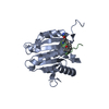
Mass: 18.015 Da / Num. of mol.: 28 / Source method: isolated from a natural source / Formula: H2O

-
Experimental details

-
Experiment

ExperimentMethod: X-RAY DIFFRACTION / Number of used crystals: 1

-
Sample preparation

CrystalDensity Matthews: 2.42 Å3/Da / Density % sol: 49.23 % / Description: NONE
Crystal growpH: 5.5
Details: 18-26% OF POLYETHYLENE GLYCOL 3350, 0.01-02M AMMONIUM SULPHATE AND 100MM BIS-TRIS AT PH 5.5

-
Data collection

DiffractionMean temperature: 100 K
Diffraction sourceSource: ROTATING ANODE / Type: BRUKER X8 PROTEUM / Wavelength: 1.5418
DetectorDetector: CCD / Date: Oct 21, 2012 / Details: MIRRORS
RadiationProtocol: SINGLE WAVELENGTH / Monochromatic (M) / Laue (L): M / Scattering type: x-ray
Radiation wavelengthWavelength: 1.5418 Å / Relative weight: 1
ReflectionResolution: 2.57→68.95 Å / Num. obs: 8036 / % possible obs: 98.8 % / Observed criterion σ(I): -3 / Redundancy: 10.15 % / Rmerge(I) obs: 0.09 / Net I/σ(I): 11.08
Reflection shellResolution: 2.57→2.63 Å / Redundancy: 2.64 % / Rmerge(I) obs: 0.46 / Mean I/σ(I) obs: 2.33 / % possible all: 83.2

-
Processing

Software
NameVersionClassification
REFMAC5.7.0032refinement
PROTEUM2data reduction
PROTEUM2data scaling
PHASERphasing
RefinementMethod to determine structure: MOLECULAR REPLACEMENT
Starting model: PDB ENTRY 4AZA
Resolution: 2.57→45.88 Å / Cor.coef. Fo:Fc: 0.876 / Cor.coef. Fo:Fc free: 0.826 / SU B: 10.265 / SU ML: 0.232 / Cross valid method: THROUGHOUT / ESU R: 0.695 / ESU R Free: 0.322 / Stereochemistry target values: MAXIMUM LIKELIHOOD
Details: HYDROGENS HAVE BEEN ADDED IN THE RIDING POSITIONS. U VALUES REFINED INDIVIDUALLY
RfactorNum. reflection% reflectionSelection details
Rfree0.26575 371 4.6 %RANDOM
Rwork0.22025 ---
obs0.22232 7640 98.88 %-
Solvent computationIon probe radii: 0.8 Å / Shrinkage radii: 0.8 Å / VDW probe radii: 1.2 Å / Solvent model: MASK
Displacement parametersBiso mean: 15.144 Å2
Baniso -1Baniso -2Baniso -3
1--0.36 Å20 Å20 Å2
2--0.4 Å20 Å2
3----0.04 Å2
Refinement stepCycle: LAST / Resolution: 2.57→45.88 Å
ProteinNucleic acidLigandSolventTotal
Num. atoms1601 0 0 28 1629
Refine LS restraints
Refine-IDTypeDev idealDev ideal targetNumber
X-RAY DIFFRACTIONr_bond_refined_d0.0090.0191639
X-RAY DIFFRACTIONr_bond_other_d0.0030.021573
X-RAY DIFFRACTIONr_angle_refined_deg1.3551.9522204
X-RAY DIFFRACTIONr_angle_other_deg0.78133601
X-RAY DIFFRACTIONr_dihedral_angle_1_deg6.9325186
X-RAY DIFFRACTIONr_dihedral_angle_2_deg38.07123.29382
X-RAY DIFFRACTIONr_dihedral_angle_3_deg16.82715292
X-RAY DIFFRACTIONr_dihedral_angle_4_deg19.8581514
X-RAY DIFFRACTIONr_chiral_restr0.0770.2233
X-RAY DIFFRACTIONr_gen_planes_refined0.0040.021801
X-RAY DIFFRACTIONr_gen_planes_other0.0010.02407
X-RAY DIFFRACTIONr_nbd_refined
X-RAY DIFFRACTIONr_nbd_other
X-RAY DIFFRACTIONr_nbtor_refined
X-RAY DIFFRACTIONr_nbtor_other
X-RAY DIFFRACTIONr_xyhbond_nbd_refined
X-RAY DIFFRACTIONr_xyhbond_nbd_other
X-RAY DIFFRACTIONr_metal_ion_refined
X-RAY DIFFRACTIONr_metal_ion_other
X-RAY DIFFRACTIONr_symmetry_vdw_refined
X-RAY DIFFRACTIONr_symmetry_vdw_other
X-RAY DIFFRACTIONr_symmetry_hbond_refined
X-RAY DIFFRACTIONr_symmetry_hbond_other
X-RAY DIFFRACTIONr_symmetry_metal_ion_refined
X-RAY DIFFRACTIONr_symmetry_metal_ion_other
X-RAY DIFFRACTIONr_mcbond_it
X-RAY DIFFRACTIONr_mcbond_other
X-RAY DIFFRACTIONr_mcangle_it
X-RAY DIFFRACTIONr_mcangle_other
X-RAY DIFFRACTIONr_scbond_it
X-RAY DIFFRACTIONr_scbond_other
X-RAY DIFFRACTIONr_scangle_it
X-RAY DIFFRACTIONr_scangle_other
X-RAY DIFFRACTIONr_long_range_B_refined
X-RAY DIFFRACTIONr_long_range_B_other
X-RAY DIFFRACTIONr_rigid_bond_restr
X-RAY DIFFRACTIONr_sphericity_free
X-RAY DIFFRACTIONr_sphericity_bonded
LS refinement shellResolution: 2.57→2.637 Å / Total num. of bins used: 20
RfactorNum. reflection% reflection
Rfree0.392 28 -
Rwork0.282 478 -
obs--86.79 %

+
About Yorodumi

-
News

-
Feb 9, 2022. New format data for meta-information of EMDB entries

New format data for meta-information of EMDB entries

  • Version 3 of the EMDB header file is now the official format.
  • The previous official version 1.9 will be removed from the archive.

Related info.:EMDB header

External links:wwPDB to switch to version 3 of the EMDB data model

-
Aug 12, 2020. Covid-19 info

Covid-19 info

URL: https://pdbjlc1.pdbj.org/emnavi/covid19.php

New page: Covid-19 featured information page in EM Navigator.

Related info.:Covid-19 info / Mar 5, 2020. Novel coronavirus structure data

+
Mar 5, 2020. Novel coronavirus structure data

Novel coronavirus structure data

Related info.:Yorodumi Speices / Aug 12, 2020. Covid-19 info

External links:COVID-19 featured content - PDBj / Molecule of the Month (242):Coronavirus Proteases

+
Jan 31, 2019. EMDB accession codes are about to change! (news from PDBe EMDB page)

EMDB accession codes are about to change! (news from PDBe EMDB page)

  • The allocation of 4 digits for EMDB accession codes will soon come to an end. Whilst these codes will remain in use, new EMDB accession codes will include an additional digit and will expand incrementally as the available range of codes is exhausted. The current 4-digit format prefixed with “EMD-” (i.e. EMD-XXXX) will advance to a 5-digit format (i.e. EMD-XXXXX), and so on. It is currently estimated that the 4-digit codes will be depleted around Spring 2019, at which point the 5-digit format will come into force.
  • The EM Navigator/Yorodumi systems omit the EMD- prefix.

Related info.:Q: What is EMD? / ID/Accession-code notation in Yorodumi/EM Navigator

External links:EMDB Accession Codes are Changing Soon! / Contact to PDBj

+
Jul 12, 2017. Major update of PDB

Major update of PDB

  • wwPDB released updated PDB data conforming to the new PDBx/mmCIF dictionary.
  • This is a major update changing the version number from 4 to 5, and with Remediation, in which all the entries are updated.
  • In this update, many items about electron microscopy experimental information are reorganized (e.g. em_software).
  • Now, EM Navigator and Yorodumi are based on the updated data.

External links:wwPDB Remediation / Enriched Model Files Conforming to OneDep Data Standards Now Available in the PDB FTP Archive

-
Yorodumi

Thousand views of thousand structures

  • Yorodumi is a browser for structure data from EMDB, PDB, SASBDB, etc.
  • This page is also the successor to EM Navigator detail page, and also detail information page/front-end page for Omokage search.
  • The word "yorodu" (or yorozu) is an old Japanese word meaning "ten thousand". "mi" (miru) is to see.

Related info.:EMDB / PDB / SASBDB / Comparison of 3 databanks / Yorodumi Search / Aug 31, 2016. New EM Navigator & Yorodumi / Yorodumi Papers / Jmol/JSmol / Function and homology information / Changes in new EM Navigator and Yorodumi

Read more