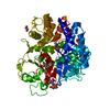
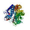
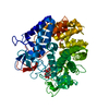
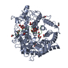
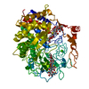
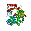
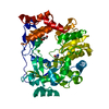
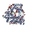
Entry Database : PDB / ID : 2v8iTitle Structure of a Family 2 Pectate Lyase in a Native Form PECTATE LYASE Keywords / / / / Function / homology Function Domain/homology Component
/ / / / / / / / / / / / / / / / / / / / / / / / / Biological species YERSINIA ENTEROCOLITICA (bacteria)Method / / Resolution : 1.5 Å Authors Abbott, D.W. / Boraston, A.B. Journal : J.Biol.Chem. / Year : 2007Title : A Family 2 Pectate Lyase Displays a Rare Fold and Transition Metal-Assisted -Elimination.Authors : Abbott, D.W. / Boraston, A.B. History Deposition Aug 8, 2007 Deposition site / Processing site Revision 1.0 Sep 18, 2007 Provider / Type Revision 1.1 May 8, 2011 Group Revision 1.2 Jul 13, 2011 Group Revision 1.3 May 8, 2024 Group / Database references / OtherCategory chem_comp_atom / chem_comp_bond ... chem_comp_atom / chem_comp_bond / database_2 / pdbx_database_status Item / _database_2.pdbx_database_accession / _pdbx_database_status.status_code_sf
Show all Show less
 Movie
Movie Controller


 Open data
 Basic information
 Components
 Keywords
 Function and homology information
 YERSINIA ENTEROCOLITICA (bacteria)
 X-RAY DIFFRACTION /
  Authors
 Citation
 Structure visualization
 Downloads & links
 Other downloads
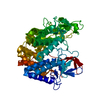


 PDBj
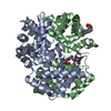
PDBj Assembly
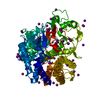

 Mass: 126.904 Da / Num. of mol.: 51 / Source method: obtained synthetically / Formula: I
 Mass: 18.015 Da / Num. of mol.: 759 / Source method: isolated from a natural source / Formula: H2O
 Sample preparation
 Processing