 Movie
Movie Controller

[English] 日本語
 Yorodumi
- PDB-2v2f: Crystal structure of PBP1a from drug-resistant strain 5204 from S... -
+ Open data

- Basic information
| Entry | Database: PDB / ID: 2v2f | ||||||
|---|---|---|---|---|---|---|---|
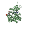
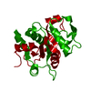
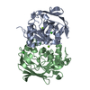
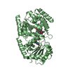
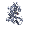
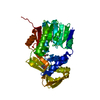
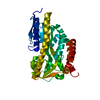
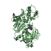
| Title | Crystal structure of PBP1a from drug-resistant strain 5204 from Streptococcus pneumoniae | ||||||
 Components Components | (PENICILLIN BINDING PROTEIN 1A) x 2 | ||||||
 Keywords Keywords | TRANSFERASE / TRANSPEPTIDASE ACTIVITY / PEPTIDOGLYCAN SYNTHESIS / HYDROLASE | ||||||
| Function / homology |  Function and homology information Function and homology informationpeptidoglycan glycosyltransferase / peptidoglycan glycosyltransferase activity / serine-type D-Ala-D-Ala carboxypeptidase / serine-type D-Ala-D-Ala carboxypeptidase activity / penicillin binding / peptidoglycan biosynthetic process / cell wall organization / regulation of cell shape / outer membrane-bounded periplasmic space / response to antibiotic ...peptidoglycan glycosyltransferase / peptidoglycan glycosyltransferase activity / serine-type D-Ala-D-Ala carboxypeptidase / serine-type D-Ala-D-Ala carboxypeptidase activity / penicillin binding / peptidoglycan biosynthetic process / cell wall organization / regulation of cell shape / outer membrane-bounded periplasmic space / response to antibiotic / proteolysis / extracellular region Similarity search - Function | ||||||
| Biological species |   STREPTOCOCCUS PNEUMONIAE (bacteria) STREPTOCOCCUS PNEUMONIAE (bacteria) | ||||||
| Method |  X-RAY DIFFRACTION / SYNCHROTRON / MOLECULAR REPLACEMENT / Resolution: 1.9 Å X-RAY DIFFRACTION / SYNCHROTRON / MOLECULAR REPLACEMENT / Resolution: 1.9 Å | ||||||
 Authors Authors | Job, V. / Carapito, R. / Vernet, T. / Dideberg, O. / Dessen, A. / Zapun, A. | ||||||
 Citation Citation | Journal: J.Biol.Chem. / Year: 2008 Title: Common Alterations in Pbp1A from Resistant Streptococcus Pneumoniae Decrease its Reactivity Toward {Beta}-Lactams: Structural Insights. Authors: Job, V. / Carapito, R. / Vernet, T. / Dessen, A. / Zapun, A. | ||||||
| History |
|
- Structure visualization
| Structure viewer | Molecule: MolmilJmol/JSmol |
|---|
- Downloads & links
-Download
| PDBx/mmCIF format | 2v2f.cif.gz | 94.1 KB | Display | PDBx/mmCIF format |
|---|---|---|---|---|
| PDB format | pdb2v2f.ent.gz | 68.9 KB | Display | PDB format |
| PDBx/mmJSON format | 2v2f.json.gz | Tree view | PDBx/mmJSON format | |
| Others |  Other downloads Other downloads |
-Validation report
| Arichive directory | https://data.pdbj.org/pub/pdb/validation_reports/v2/2v2fftp://data.pdbj.org/pub/pdb/validation_reports/v2/2v2f | HTTPS FTP |
|---|
-Related structure data
| Related structure data |  2cw6S S: Starting model for refinement |
|---|---|
| Similar structure data |

-Links
 PDBj
PDBj
- Assembly
| Deposited unit | 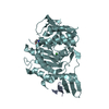
| ||||||||
|---|---|---|---|---|---|---|---|---|---|
| 1 |
| ||||||||
| Unit cell |
|
-Components
| #1: Protein/peptide | Mass: 2639.935 Da / Num. of mol.: 1 / Fragment: GLYCOSYLTRASFERASE DOMAIN, RESIDUES 47-70 Source method: isolated from a genetically manipulated source Source: (gene. exp.) STREPTOCOCCUS PNEUMONIAE (bacteria) / Strain: 5204 / Production host: References: UniProt: Q9RET4, peptidoglycan glycosyltransferase |
|---|---|
| #2: Protein | Mass: 43432.023 Da / Num. of mol.: 1 Fragment: TRANSPEPTIDASE DOMAIN, GLYCOSYLTRASFERASE DOMAIN, RESIDUES 264-653 Source method: isolated from a genetically manipulated source Source: (gene. exp.) STREPTOCOCCUS PNEUMONIAE (bacteria) / Strain: 5204 / Production host: References: UniProt: Q9RET4, serine-type D-Ala-D-Ala carboxypeptidase |
| #3: Chemical | ChemComp-BA /   Mass: 137.327 Da / Num. of mol.: 1 / Source method: obtained synthetically / Formula: Ba Mass: 137.327 Da / Num. of mol.: 1 / Source method: obtained synthetically / Formula: Ba |
| #4: Chemical | ChemComp-MES /   Mass: 195.237 Da / Num. of mol.: 1 / Source method: obtained synthetically / Formula: C6H13NO4S / Comment: pH buffer*YM Mass: 195.237 Da / Num. of mol.: 1 / Source method: obtained synthetically / Formula: C6H13NO4S / Comment: pH buffer*YM |
| #5: Water | ChemComp-HOH /  Mass: 18.015 Da / Num. of mol.: 203 / Source method: isolated from a natural source / Formula: H2O Mass: 18.015 Da / Num. of mol.: 203 / Source method: isolated from a natural source / Formula: H2O |
-Experimental details
-Experiment
| Experiment | Method: X-RAY DIFFRACTION / Number of used crystals: 1 |
|---|
- Sample preparation
| Crystal | Density Matthews: 2.3 Å3/Da / Density % sol: 45.9 % / Description: NONE |
|---|---|
| Crystal grow | pH: 6 / Details: 0.1M MES PH6, 21% PEG6000, 17MM BACL2 |
-Data collection
| Diffraction | Mean temperature: 287 K |
|---|---|
| Diffraction source | Source: SYNCHROTRON / Site: ESRF  / Beamline: ID14-3 / Wavelength: 0.931 / Beamline: ID14-3 / Wavelength: 0.931 |
| Detector | Type: ADSC CCD / Detector: CCD / Date: Nov 5, 2005 |
| Radiation | Protocol: SINGLE WAVELENGTH / Monochromatic (M) / Laue (L): M / Scattering type: x-ray |
| Radiation wavelength | Wavelength: 0.931 Å / Relative weight: 1 |
| Reflection | Resolution: 1.9→19.7 Å / Num. obs: 29802 / % possible obs: 96.1 % / Observed criterion σ(I): 0 / Redundancy: 3.77 % / Biso Wilson estimate: 18.23 Å2 / Rmerge(I) obs: 0.12 / Net I/σ(I): 11.1 |
| Reflection shell | Resolution: 1.9→2.06 Å / Redundancy: 3.86 % / Rmerge(I) obs: 0.37 / Mean I/σ(I) obs: 4 / % possible all: 93.2 |
- Processing
| Software |
| ||||||||||||||||||||||||||||||||||||||||||||||||||||||||||||||||||||||||||||||||||||||||||||||||||||||||||||||||||||||||||||||||||||||||||||||||||||||||||||||||||||||||||||||||||||||
|---|---|---|---|---|---|---|---|---|---|---|---|---|---|---|---|---|---|---|---|---|---|---|---|---|---|---|---|---|---|---|---|---|---|---|---|---|---|---|---|---|---|---|---|---|---|---|---|---|---|---|---|---|---|---|---|---|---|---|---|---|---|---|---|---|---|---|---|---|---|---|---|---|---|---|---|---|---|---|---|---|---|---|---|---|---|---|---|---|---|---|---|---|---|---|---|---|---|---|---|---|---|---|---|---|---|---|---|---|---|---|---|---|---|---|---|---|---|---|---|---|---|---|---|---|---|---|---|---|---|---|---|---|---|---|---|---|---|---|---|---|---|---|---|---|---|---|---|---|---|---|---|---|---|---|---|---|---|---|---|---|---|---|---|---|---|---|---|---|---|---|---|---|---|---|---|---|---|---|---|---|---|---|---|
| Refinement | Method to determine structure: MOLECULAR REPLACEMENT Starting model: PDB ENTRY 2CW6 Resolution: 1.9→19.74 Å / Cor.coef. Fo:Fc: 0.911 / Cor.coef. Fo:Fc free: 0.876 / Cross valid method: THROUGHOUT / ESU R: 0.195 / ESU R Free: 0.166 / Stereochemistry target values: MAXIMUM LIKELIHOOD / Details: HYDROGENS HAVE BEEN ADDED IN THE RIDING POSITIONS.
| ||||||||||||||||||||||||||||||||||||||||||||||||||||||||||||||||||||||||||||||||||||||||||||||||||||||||||||||||||||||||||||||||||||||||||||||||||||||||||||||||||||||||||||||||||||||
| Solvent computation | Ion probe radii: 0.8 Å / Shrinkage radii: 0.8 Å / VDW probe radii: 1.2 Å / Solvent model: MASK | ||||||||||||||||||||||||||||||||||||||||||||||||||||||||||||||||||||||||||||||||||||||||||||||||||||||||||||||||||||||||||||||||||||||||||||||||||||||||||||||||||||||||||||||||||||||
| Displacement parameters | Biso mean: 13.11 Å2
| ||||||||||||||||||||||||||||||||||||||||||||||||||||||||||||||||||||||||||||||||||||||||||||||||||||||||||||||||||||||||||||||||||||||||||||||||||||||||||||||||||||||||||||||||||||||
| Refinement step | Cycle: LAST / Resolution: 1.9→19.74 Å
| ||||||||||||||||||||||||||||||||||||||||||||||||||||||||||||||||||||||||||||||||||||||||||||||||||||||||||||||||||||||||||||||||||||||||||||||||||||||||||||||||||||||||||||||||||||||
| Refine LS restraints |
|