 Movie
Movie Controller

[English] 日本語
 Yorodumi
- PDB-2v1r: Yeast Pex13 SH3 domain complexed with a peptide from Pex14 at 2.1... -
+ Open data

- Basic information
| Entry | Database: PDB / ID: 2v1r | ||||||
|---|---|---|---|---|---|---|---|
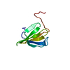
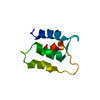
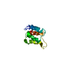
| Title | Yeast Pex13 SH3 domain complexed with a peptide from Pex14 at 2.1 A resolution | ||||||
 Components Components |
| ||||||
 Keywords Keywords | PROTEIN TRANSPORT / TRANSLOCATION / TRANSMEMBRANE / PEPTIDE COMPLEX / STRUCTURAL GENOMICS / PEROXISOME | ||||||
| Function / homology |  Function and homology information Function and homology informationperoxisomal importomer complex / Class I peroxisomal membrane protein import / protein import into peroxisome matrix, docking / Peroxisomal protein import / : / peroxisomal membrane / transmembrane protein transporter activity / protein-macromolecule adaptor activity / signaling receptor binding Similarity search - Function | ||||||
| Biological species |  | ||||||
| Method |  X-RAY DIFFRACTION / SYNCHROTRON / MOLECULAR REPLACEMENT / Resolution: 2.1 Å X-RAY DIFFRACTION / SYNCHROTRON / MOLECULAR REPLACEMENT / Resolution: 2.1 Å | ||||||
 Authors Authors | Kursula, I. / Kursula, P. / Lehmann, F. / Zou, P. / Song, Y.H. / Wilmanns, M. | ||||||
 Citation Citation | Journal: To be Published Title: Structural Genomics of Yeast SH3 Domains Authors: Kursula, P. / Kursula, I. / Pinotsis, N. / Lehmann, F. / Zou, P. / Song, Y.H. / Wilmanns, M. | ||||||
| History |
|
- Structure visualization
| Structure viewer | Molecule: MolmilJmol/JSmol |
|---|
- Downloads & links
-Download
| PDBx/mmCIF format | 2v1r.cif.gz | 45.8 KB | Display | PDBx/mmCIF format |
|---|---|---|---|---|
| PDB format | pdb2v1r.ent.gz | 32.7 KB | Display | PDB format |
| PDBx/mmJSON format | 2v1r.json.gz | Tree view | PDBx/mmJSON format | |
| Others |  Other downloads Other downloads |
-Validation report
| Arichive directory | https://data.pdbj.org/pub/pdb/validation_reports/v1/2v1rftp://data.pdbj.org/pub/pdb/validation_reports/v1/2v1r | HTTPS FTP |
|---|
-Related structure data
| Related structure data |  2v1qC  2vknC  1n5zS S: Starting model for refinement C: citing same article ( |
|---|---|
| Similar structure data |


-Links
 PDBj
PDBj

- Assembly
| Deposited unit | 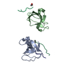
| ||||||||
|---|---|---|---|---|---|---|---|---|---|
| 1 | 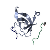
| ||||||||
| 2 | 
| ||||||||
| Unit cell |
|
-Components
| #1: Protein | Mass: 9316.717 Da / Num. of mol.: 2 / Fragment: SH3 DOMAIN, RESIDUES 299-374 Source method: isolated from a genetically manipulated source Source: (gene. exp.) Production host:  #2: Protein/peptide | Mass: 1721.952 Da / Num. of mol.: 3 / Fragment: SH3 DOMAIN BINDING SEGMENT, RESIDUES 83-96 / Source method: obtained synthetically / Source: (synth.) #3: Water | ChemComp-HOH / |  Mass: 18.015 Da / Num. of mol.: 105 / Source method: isolated from a natural source / Formula: H2O Mass: 18.015 Da / Num. of mol.: 105 / Source method: isolated from a natural source / Formula: H2OHas protein modification | Y | |
|---|
-Experimental details
-Experiment
| Experiment | Method: X-RAY DIFFRACTION / Number of used crystals: 1 |
|---|
- Sample preparation
| Crystal | Density Matthews: 2.3 Å3/Da / Density % sol: 46.14 % / Description: NONE |
|---|
-Data collection
| Diffraction | Mean temperature: 100 K |
|---|---|
| Diffraction source | Source: SYNCHROTRON / Site: EMBL/DESY, HAMBURG  / Beamline: X11 / Wavelength: 0.81 / Beamline: X11 / Wavelength: 0.81 |
| Detector | Type: MARRESEARCH / Detector: CCD |
| Radiation | Protocol: SINGLE WAVELENGTH / Monochromatic (M) / Laue (L): M / Scattering type: x-ray |
| Radiation wavelength | Wavelength: 0.81 Å / Relative weight: 1 |
| Reflection | Resolution: 2.11→20 Å / Num. obs: 9925 / % possible obs: 93.7 % / Observed criterion σ(I): -3 / Redundancy: 2 % / Rmerge(I) obs: 0.06 / Net I/σ(I): 9.5 |
| Reflection shell | Resolution: 2.11→2.25 Å / Redundancy: 1.7 % / Rmerge(I) obs: 0.39 / Mean I/σ(I) obs: 2.6 / % possible all: 87 |
- Processing
| Software |
| ||||||||||||||||
|---|---|---|---|---|---|---|---|---|---|---|---|---|---|---|---|---|---|
| Refinement | Method to determine structure: MOLECULAR REPLACEMENT Starting model: PDB ENTRY 1N5Z Resolution: 2.1→20 Å
| ||||||||||||||||
| Refinement step | Cycle: LAST / Resolution: 2.1→20 Å
|
