- PDB-2v0p: The Structure of Tap42 Alpha4 Subunit -
+
Open data
ID or keywords:
Loading...
-
Basic information
Entry
Database: PDB / ID: 2v0p
Title
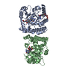
The Structure of Tap42 Alpha4 Subunit
Components
TYPE 2A PHOSPHATASE-ASSOCIATED PROTEIN 42
Keywords
HYDROLASE INHIBITOR / PHOSPHORYLATION / SIGNAL TRANSDUCTION INHIBITOR
Function / homology
Function and homology information
regulation of dephosphorylation / protein phosphatase type 2A complex / regulation of TORC1 signaling / positive regulation of transcription by RNA polymerase I / TOR signaling / negative regulation of signal transduction / protein phosphatase 2A binding / cytosol Similarity search - Function
TAP42-like family / TAP46-like protein / TAP42/TAP46-like superfamily / TAP42-like family / Serine Threonine Protein Phosphatase 5, Tetratricopeptide repeat / Alpha Horseshoe / Mainly Alpha Similarity search - Domain/homology
Mass: 18.015 Da / Num. of mol.: 253 / Source method: isolated from a natural source / Formula: H2O
Has protein modification
Y
-
Experimental details
-
Experiment
Experiment
Method: X-RAY DIFFRACTION / Number of used crystals: 1
-
Sample preparation
Crystal
Density Matthews: 2.7 Å3/Da / Density % sol: 54 %
Crystal grow
Temperature: 293 K / Method: vapor diffusion, hanging drop / pH: 6.5 Details: THE PROTEIN WAS CONCENTRATED TO 10 MG/ML BEFORE CRYSTALLIZATION. CRYSTALS WERE GROWN AT 20C USING THE HANGING DROP METHOD. ONE MICROLITRE OF PROTEIN WAS MIXED WITH AN EQUAL VOLUME OF ...Details: THE PROTEIN WAS CONCENTRATED TO 10 MG/ML BEFORE CRYSTALLIZATION. CRYSTALS WERE GROWN AT 20C USING THE HANGING DROP METHOD. ONE MICROLITRE OF PROTEIN WAS MIXED WITH AN EQUAL VOLUME OF CRYSTALLIZATION BUFFER: 20% (W/V) PEG 8000, 0.1 M SODIUM CACODYLATE PH 6.5, 0.2 M MAGNESIUM ACETATE AND 2MM DTT. CRYSTALS WERE OPTIMISED BY SEEDING.
 Movie
Movie Controller


 Open data
 Basic information
 Components
 Keywords
 Function and homology information
 X-RAY DIFFRACTION /
  Authors
 Citation
 Structure visualization
 Downloads & links
 Other downloads


 PDBj
PDBj Assembly


 Mass: 65.409 Da / Num. of mol.: 4 / Source method: obtained synthetically / Formula: Zn
 Mass: 18.015 Da / Num. of mol.: 253 / Source method: isolated from a natural source / Formula: H2O
 Sample preparation
 / Beamline: BM14 / Wavelength: 0.9775
  Processing