 Movie
Movie Controller

[English] 日本語
 Yorodumi
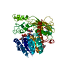
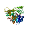
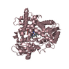
- PDB-2uyt: Structure of L-rhamnulose kinase in complex with ADP and beta-L- ... -
+ Open data

- Basic information
| Entry | Database: PDB / ID: 2uyt | ||||||
|---|---|---|---|---|---|---|---|
| Title | Structure of L-rhamnulose kinase in complex with ADP and beta-L- rhamnulose. | ||||||
 Components Components | RHAMNULOKINASE | ||||||
 Keywords Keywords | TRANSFERASE / RHAMNOSE DEGRADATION / IN-LINE PHOSPHORYL TRANSFER / HEXOKINASE-HSP70-ACTIN SUPERFAMILY / L-RHAMNULOSE KINASE / RHAMNOSE METABOLISM / KINASE / INDUCED FIT / RARE SUGARS | ||||||
| Function / homology |  Function and homology information Function and homology informationrhamnulokinase / rhamnulokinase activity / rhamnose catabolic process / glycerol kinase activity / glycerol metabolic process / ATP binding / cytosol Similarity search - Function | ||||||
| Biological species |  | ||||||
| Method |  X-RAY DIFFRACTION / SYNCHROTRON / MOLECULAR REPLACEMENT / Resolution: 1.55 Å X-RAY DIFFRACTION / SYNCHROTRON / MOLECULAR REPLACEMENT / Resolution: 1.55 Å | ||||||
 Authors Authors | Grueninger, D. / Schulz, G.E. | ||||||
 Citation Citation | Journal: FEBS Lett. / Year: 2007 Title: Substrate Spectrum of L-Rhamnulose Kinase Related to Models Derived from Two Ternary Complex Structures. Authors: Grueninger, D. / Schulz, G.E. #1: Journal: J.Mol.Biol. / Year: 2006Title: Structure and Reaction Mechanism of L-Rhamnulose Kinase from Escherichia Coli Authors: Grueninger, D. / Schulz, G.E. | ||||||
| History |
|
- Structure visualization
| Structure viewer | Molecule: MolmilJmol/JSmol |
|---|
- Downloads & links
-Download
| PDBx/mmCIF format | 2uyt.cif.gz | 114.8 KB | Display | PDBx/mmCIF format |
|---|---|---|---|---|
| PDB format | pdb2uyt.ent.gz | 87.7 KB | Display | PDB format |
| PDBx/mmJSON format | 2uyt.json.gz | Tree view | PDBx/mmJSON format | |
| Others |  Other downloads Other downloads |
-Validation report
| Arichive directory | https://data.pdbj.org/pub/pdb/validation_reports/uy/2uytftp://data.pdbj.org/pub/pdb/validation_reports/uy/2uyt | HTTPS FTP |
|---|
-Related structure data
| Related structure data |  2cglS S: Starting model for refinement |
|---|---|
| Similar structure data |


-Links
 PDBj
PDBj- Assembly
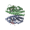
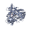
| Deposited unit | 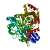
| ||||||||
|---|---|---|---|---|---|---|---|---|---|
| 1 |
| ||||||||
| Unit cell |
|
-Components
| #1: Protein | Mass: 53954.715 Da / Num. of mol.: 1 / Mutation: YES Source method: isolated from a genetically manipulated source Source: (gene. exp.) References: UniProt: Q8X899, UniProt: Q1R415*PLUS, rhamnulokinase |
|---|---|
| #2: Sugar | ChemComp-LRH / 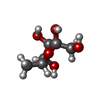  Type: L-saccharide, beta linking / Mass: 164.156 Da / Num. of mol.: 1 / Source method: obtained synthetically / Formula: C6H12O5 Type: L-saccharide, beta linking / Mass: 164.156 Da / Num. of mol.: 1 / Source method: obtained synthetically / Formula: C6H12O5 |
| #3: Chemical | ChemComp-ADP /   Mass: 427.201 Da / Num. of mol.: 1 / Source method: obtained synthetically / Formula: C10H15N5O10P2 / Comment: ADP, energy-carrying molecule*YM Mass: 427.201 Da / Num. of mol.: 1 / Source method: obtained synthetically / Formula: C10H15N5O10P2 / Comment: ADP, energy-carrying molecule*YM |
| #4: Water | ChemComp-HOH /  Mass: 18.015 Da / Num. of mol.: 322 / Source method: isolated from a natural source / Formula: H2O Mass: 18.015 Da / Num. of mol.: 322 / Source method: isolated from a natural source / Formula: H2O |
| Compound details | ENGINEERED RESIDUE IN CHAIN A, GLU 69 TO ALA ENGINEERED RESIDUE IN CHAIN A, GLU 70 TO ALA ...ENGINEERED |
| Has protein modification | Y |
| Sequence details | THE SEQUENCE DISCREPANCIES ARE BECAUSE THE DATABASE SEQUENCE IS FROM E.COLI STRAIN O157-H7. THE ...THE SEQUENCE DISCREPANC |
-Experimental details
-Experiment
| Experiment | Method: X-RAY DIFFRACTION |
|---|
- Sample preparation
| Crystal | Density Matthews: 1.9 Å3/Da / Density % sol: 35 % |
|---|---|
| Crystal grow | Details: 17% (W/V) PEG 8000, 0.12 M LICL, 20 MM ADP, 20 MM L-RHAMNULOSE-1-PHOSPHATE |
-Data collection
| Diffraction | Mean temperature: 100 K |
|---|---|
| Diffraction source | Source: SYNCHROTRON / Site: SLS  / Beamline: X06SA / Wavelength: 0.91841 / Beamline: X06SA / Wavelength: 0.91841 |
| Detector | Type: MARRESEARCH / Detector: CCD |
| Radiation | Protocol: SINGLE WAVELENGTH / Monochromatic (M) / Laue (L): M / Scattering type: x-ray |
| Radiation wavelength | Wavelength: 0.91841 Å / Relative weight: 1 |
| Reflection | Resolution: 1.55→80 Å / Num. obs: 60401 / % possible obs: 98.6 % / Observed criterion σ(I): 4.7 / Redundancy: 6.9 % / Rmerge(I) obs: 0.05 / Net I/σ(I): 23.2 |
| Reflection shell | Resolution: 1.55→1.62 Å / Redundancy: 5.5 % / Rmerge(I) obs: 0.37 / Mean I/σ(I) obs: 4.7 / % possible all: 89.4 |
- Processing
| Software |
| ||||||||||||||||||||||||||||||||||||||||||||||||||||||||||||||||||||||||||||||||||||||||||||||||||||||||||||||||||||||||||||||||||||||||||||||||||||||||||||||||||||||||||||||||||||||
|---|---|---|---|---|---|---|---|---|---|---|---|---|---|---|---|---|---|---|---|---|---|---|---|---|---|---|---|---|---|---|---|---|---|---|---|---|---|---|---|---|---|---|---|---|---|---|---|---|---|---|---|---|---|---|---|---|---|---|---|---|---|---|---|---|---|---|---|---|---|---|---|---|---|---|---|---|---|---|---|---|---|---|---|---|---|---|---|---|---|---|---|---|---|---|---|---|---|---|---|---|---|---|---|---|---|---|---|---|---|---|---|---|---|---|---|---|---|---|---|---|---|---|---|---|---|---|---|---|---|---|---|---|---|---|---|---|---|---|---|---|---|---|---|---|---|---|---|---|---|---|---|---|---|---|---|---|---|---|---|---|---|---|---|---|---|---|---|---|---|---|---|---|---|---|---|---|---|---|---|---|---|---|---|
| Refinement | Method to determine structure: MOLECULAR REPLACEMENT Starting model: PDB ENTRY 2CGL Resolution: 1.55→79.31 Å / Cor.coef. Fo:Fc: 0.959 / Cor.coef. Fo:Fc free: 0.944 / SU B: 3.162 / SU ML: 0.061 / TLS residual ADP flag: LIKELY RESIDUAL / Cross valid method: THROUGHOUT / ESU R: 0.091 / ESU R Free: 0.089 / Stereochemistry target values: MAXIMUM LIKELIHOOD / Details: HYDROGENS HAVE BEEN ADDED IN THE RIDING POSITIONS.
| ||||||||||||||||||||||||||||||||||||||||||||||||||||||||||||||||||||||||||||||||||||||||||||||||||||||||||||||||||||||||||||||||||||||||||||||||||||||||||||||||||||||||||||||||||||||
| Solvent computation | Ion probe radii: 0.8 Å / Shrinkage radii: 0.8 Å / VDW probe radii: 1.4 Å / Solvent model: MASK | ||||||||||||||||||||||||||||||||||||||||||||||||||||||||||||||||||||||||||||||||||||||||||||||||||||||||||||||||||||||||||||||||||||||||||||||||||||||||||||||||||||||||||||||||||||||
| Displacement parameters | Biso mean: 14.5 Å2
| ||||||||||||||||||||||||||||||||||||||||||||||||||||||||||||||||||||||||||||||||||||||||||||||||||||||||||||||||||||||||||||||||||||||||||||||||||||||||||||||||||||||||||||||||||||||
| Refinement step | Cycle: LAST / Resolution: 1.55→79.31 Å
| ||||||||||||||||||||||||||||||||||||||||||||||||||||||||||||||||||||||||||||||||||||||||||||||||||||||||||||||||||||||||||||||||||||||||||||||||||||||||||||||||||||||||||||||||||||||
| Refine LS restraints |
|