 Movie
Movie Controller

[English] 日本語
 Yorodumi
- PDB-2cgk: Crystal Structure of L-rhamnulose kinase from Escherichia coli in... -
+ Open data

- Basic information
| Entry | Database: PDB / ID: 2cgk | ||||||
|---|---|---|---|---|---|---|---|
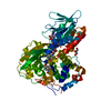
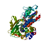
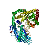
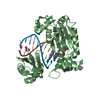
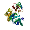
| Title | Crystal Structure of L-rhamnulose kinase from Escherichia coli in an open uncomplexed conformation. | ||||||
 Components Components | L-RHAMNULOSE KINASE | ||||||
 Keywords Keywords | TRANSFERASE / L-RHAMNULOSE KINASE / RHAMNOSE DEGRADATION / HEXOKINASE-HSP70- ACTIN SUPERFAMILY / INDUCED FIT / IN-LINE PHOSPHORYL TRANSFER / KINASE / RHAMNOSE METABOLISM | ||||||
| Function / homology |  Function and homology information Function and homology informationrhamnulokinase / rhamnulokinase activity / rhamnose catabolic process / glycerol kinase activity / glycerol metabolic process / ATP binding / cytosol Similarity search - Function | ||||||
| Biological species |  | ||||||
| Method |  X-RAY DIFFRACTION / SYNCHROTRON / MOLECULAR REPLACEMENT / Resolution: 2.46 Å X-RAY DIFFRACTION / SYNCHROTRON / MOLECULAR REPLACEMENT / Resolution: 2.46 Å | ||||||
 Authors Authors | Grueninger, D. / Schulz, G.E. | ||||||
 Citation Citation | Journal: J.Mol.Biol. / Year: 2006 Title: Structure and Reaction Mechanism of L-Rhamnulose Kinase from Escherichia Coli. Authors: Grueninger, D. / Schulz, G.E. | ||||||
| History |
| ||||||
| Remark 700 | SHEET THE SHEET STRUCTURE OF THIS MOLECULE IS BIFURCATED. IN ORDER TO REPRESENT THIS FEATURE IN ... SHEET THE SHEET STRUCTURE OF THIS MOLECULE IS BIFURCATED. IN ORDER TO REPRESENT THIS FEATURE IN THE SHEET RECORDS BELOW, TWO SHEETS ARE DEFINED. |
- Structure visualization
| Structure viewer | Molecule: MolmilJmol/JSmol |
|---|
- Downloads & links
-Download
| PDBx/mmCIF format | 2cgk.cif.gz | 191.4 KB | Display | PDBx/mmCIF format |
|---|---|---|---|---|
| PDB format | pdb2cgk.ent.gz | 154.1 KB | Display | PDB format |
| PDBx/mmJSON format | 2cgk.json.gz | Tree view | PDBx/mmJSON format | |
| Others |  Other downloads Other downloads |
-Validation report
| Arichive directory | https://data.pdbj.org/pub/pdb/validation_reports/cg/2cgkftp://data.pdbj.org/pub/pdb/validation_reports/cg/2cgk | HTTPS FTP |
|---|
-Related structure data
| Related structure data |  2cgjC  2cglSC C: citing same article ( S: Starting model for refinement |
|---|---|
| Similar structure data |


-Links
 PDBj
PDBj- Assembly
| Deposited unit | 
| ||||||||
|---|---|---|---|---|---|---|---|---|---|
| 1 | 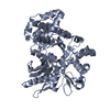
| ||||||||
| 2 | 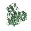
| ||||||||
| Unit cell |
| ||||||||
| Noncrystallographic symmetry (NCS) | NCS oper: (Code: given Matrix: (0.99999, 0.00391, -0.00275), Vector: |
-Components
| #1: Protein | Mass: 53954.715 Da / Num. of mol.: 2 / Mutation: YES Source method: isolated from a genetically manipulated source Source: (gene. exp.) References: UniProt: Q8X899, UniProt: P32171*PLUS, rhamnulokinase #2: Water | ChemComp-HOH / |  Mass: 18.015 Da / Num. of mol.: 94 / Source method: isolated from a natural source / Formula: H2O Mass: 18.015 Da / Num. of mol.: 94 / Source method: isolated from a natural source / Formula: H2OCompound details | ENGINEERED RESIDUE IN CHAIN A, GLU 69 TO ALA ENGINEERED RESIDUE IN CHAIN A, GLU 70 TO ALA ...ENGINEERED | Has protein modification | Y | Sequence details | THE SEQUENCE DISCREPANCIES ARE BECAUSE THE DATABASE SEQUENCE IS FROM E.COLI STRAIN O157-H7. THE ...THE SEQUENCE DISCREPANC | |
|---|
-Experimental details
-Experiment
| Experiment | Method: X-RAY DIFFRACTION / Number of used crystals: 1 |
|---|
- Sample preparation
| Crystal | Density Matthews: 2.1 Å3/Da / Density % sol: 40 % |
|---|---|
| Crystal grow | Details: 17 % PEG 8000, 120 MM LICL |
-Data collection
| Diffraction | Mean temperature: 100 K |
|---|---|
| Diffraction source | Source: SYNCHROTRON / Site: SLS  / Beamline: X06SA / Wavelength: 0.9184 / Beamline: X06SA / Wavelength: 0.9184 |
| Detector | Type: MARRESEARCH / Detector: CCD / Date: Sep 26, 2005 |
| Radiation | Monochromator: SI(111) / Protocol: SINGLE WAVELENGTH / Monochromatic (M) / Laue (L): M / Scattering type: x-ray |
| Radiation wavelength | Wavelength: 0.9184 Å / Relative weight: 1 |
| Reflection | Resolution: 2.46→52 Å / Num. obs: 29856 / % possible obs: 95.1 % / Observed criterion σ(I): 0 / Redundancy: 2.4 % / Rmerge(I) obs: 0.1 / Net I/σ(I): 8.3 |
| Reflection shell | Resolution: 2.46→2.54 Å / Redundancy: 2.4 % / Rmerge(I) obs: 0.42 / Mean I/σ(I) obs: 3.6 / % possible all: 97.5 |
- Processing
| Software |
| ||||||||||||||||||||||||||||||||||||||||||||||||||||||||||||
|---|---|---|---|---|---|---|---|---|---|---|---|---|---|---|---|---|---|---|---|---|---|---|---|---|---|---|---|---|---|---|---|---|---|---|---|---|---|---|---|---|---|---|---|---|---|---|---|---|---|---|---|---|---|---|---|---|---|---|---|---|---|
| Refinement | Method to determine structure: MOLECULAR REPLACEMENT Starting model: PDB ENTRY 2CGL Resolution: 2.46→44 Å / Data cutoff high absF: 10000 / Cross valid method: RMSD / σ(F): 0 Stereochemistry target values: MAXIMUM LIKELIHOOD TARGET USING AMPLITUDES Details: THE STRUCTURE WAS REFINED USING STRICT NCS.
| ||||||||||||||||||||||||||||||||||||||||||||||||||||||||||||
| Solvent computation | Solvent model: CNS BULK SOLVENT MODEL USED / Bsol: 28.1832 Å2 / ksol: 0.340036 e/Å3 | ||||||||||||||||||||||||||||||||||||||||||||||||||||||||||||
| Displacement parameters | Biso mean: 33.9 Å2
| ||||||||||||||||||||||||||||||||||||||||||||||||||||||||||||
| Refine analyze |
| ||||||||||||||||||||||||||||||||||||||||||||||||||||||||||||
| Refinement step | Cycle: LAST / Resolution: 2.46→44 Å
| ||||||||||||||||||||||||||||||||||||||||||||||||||||||||||||
| Refine LS restraints |
| ||||||||||||||||||||||||||||||||||||||||||||||||||||||||||||
| LS refinement shell | Resolution: 2.46→2.55 Å / Total num. of bins used: 10
| ||||||||||||||||||||||||||||||||||||||||||||||||||||||||||||
| Xplor file |
|
