- PDB-2uwj: Structure of the heterotrimeric complex which regulates type III ... -
+
Open data
ID or keywords:
Loading...
-
Basic information
Entry
Database: PDB / ID: 2uwj
Title
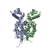
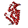
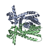
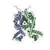
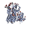
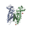
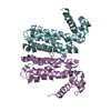
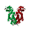
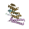
Structure of the heterotrimeric complex which regulates type III secretion needle formation
Components
TYPE III EXPORT PROTEIN PSCE
TYPE III EXPORT PROTEIN PSCF
TYPE III EXPORT PROTEIN PSCG
Keywords
CHAPERONE / VIRULENCE / CHAPERONES / COILED COIL / NEEDLE FORMATION / TYPE III SECRETION / BACTERIAL PATHOGENICITY
Function / homology
Function and homology information
type III protein secretion system complex / protein secretion by the type III secretion system / cell surface / extracellular region / cytoplasm Similarity search - Function
Type III secretion system YscG / Bacterial type III secretion system chaperone protein / Type III secretion system, secretion protein E / Type III secretion system, cytoplasmic E component of needle / Type III secretion, needle-protein-like / Type III secretion, needle-protein-like superfamily / Type III secretion needle MxiH, YscF, SsaG, EprI, PscF, EscF / Type III secretion system, needle protein / Immunoglobulin FC, subunit C / Tetratricopeptide repeat domain ...Type III secretion system YscG / Bacterial type III secretion system chaperone protein / Type III secretion system, secretion protein E / Type III secretion system, cytoplasmic E component of needle / Type III secretion, needle-protein-like / Type III secretion, needle-protein-like superfamily / Type III secretion needle MxiH, YscF, SsaG, EprI, PscF, EscF / Type III secretion system, needle protein / Immunoglobulin FC, subunit C / Tetratricopeptide repeat domain / Single alpha-helices involved in coiled-coils or other helix-helix interfaces / Serine Threonine Protein Phosphatase 5, Tetratricopeptide repeat / Alpha Horseshoe / Tetratricopeptide-like helical domain superfamily / Up-down Bundle / Mainly Alpha Similarity search - Domain/homology
NICKEL (II) ION / Type 3 secretion system needle filament protein / Type 3 secretion system chaperone PscG / Type 3 secretion system chaperone PscE Similarity search - Component
Biological species
PSEUDOMONAS AERUGINOSA (bacteria)
Method
X-RAY DIFFRACTION / SYNCHROTRON / SAD / Resolution: 2 Å
Mass: 18.015 Da / Num. of mol.: 172 / Source method: isolated from a natural source / Formula: H2O
Sequence details
N-TERMINAL GSH TAG REMAINING RESIDUES THE DNA SEQUENCE CORRESPONDING TO THE FIRST 54 RESIDUES OF ...N-TERMINAL GSH TAG REMAINING RESIDUES THE DNA SEQUENCE CORRESPONDING TO THE FIRST 54 RESIDUES OF PSCF WAS REMOVED FROM THE PSCEFG SEQUENCE.
-
Experimental details
-
Experiment
Experiment
Method: X-RAY DIFFRACTION / Number of used crystals: 1
-
Sample preparation
Crystal
Density Matthews: 3.47 Å3/Da / Density % sol: 64.57 % / Description: HEAVY ATOM SEARCH USING SHELXD
Protocol: SINGLE WAVELENGTH / Monochromatic (M) / Laue (L): M / Scattering type: x-ray
Radiation wavelength
Wavelength: 0.933 Å / Relative weight: 1
Reflection
Resolution: 2→30 Å / Num. obs: 27335 / % possible obs: 99.8 % / Observed criterion σ(I): 0 / Redundancy: 70.1 % / Biso Wilson estimate: 20.8 Å2 / Rmerge(I) obs: 0.05 / Net I/σ(I): 12.4
Reflection shell
Resolution: 2→2.07 Å / Rmerge(I) obs: 0.38 / Mean I/σ(I) obs: 3.7 / % possible all: 100
-
Processing
Software
Name
Version
Classification
CNS
1.1
refinement
DENZO
datareduction
SCALEPACK
datascaling
SHARP
phasing
Refinement
Method to determine structure: SAD / Resolution: 2→28.99 Å / Rfactor Rfree error: 0.007 / Data cutoff high absF: 2034355.35 / Isotropic thermal model: RESTRAINED / Cross valid method: THROUGHOUT / σ(F): 0 / Stereochemistry target values: MAXIMUM LIKELIHOOD / Details: PSCG FOLDS AS A TPR LIKE DOMAIN
In the structure databanks used in Yorodumi, some data are registered as the other names, "COVID-19 virus" and "2019-nCoV". Here are the details of the virus and the list of structure data.
Jan 31, 2019. EMDB accession codes are about to change! (news from PDBe EMDB page)
EMDB accession codes are about to change! (news from PDBe EMDB page)
The allocation of 4 digits for EMDB accession codes will soon come to an end. Whilst these codes will remain in use, new EMDB accession codes will include an additional digit and will expand incrementally as the available range of codes is exhausted. The current 4-digit format prefixed with “EMD-” (i.e. EMD-XXXX) will advance to a 5-digit format (i.e. EMD-XXXXX), and so on. It is currently estimated that the 4-digit codes will be depleted around Spring 2019, at which point the 5-digit format will come into force.
The EM Navigator/Yorodumi systems omit the EMD- prefix.
Related info.:Q: What is EMD? / ID/Accession-code notation in Yorodumi/EM Navigator
Yorodumi is a browser for structure data from EMDB, PDB, SASBDB, etc.
This page is also the successor to EM Navigator detail page, and also detail information page/front-end page for Omokage search.
The word "yorodu" (or yorozu) is an old Japanese word meaning "ten thousand". "mi" (miru) is to see.
Related info.:EMDB / PDB / SASBDB / Comparison of 3 databanks / Yorodumi Search / Aug 31, 2016. New EM Navigator & Yorodumi / Yorodumi Papers / Jmol/JSmol / Function and homology information / Changes in new EM Navigator and Yorodumi
 Movie
Movie Controller

 Yorodumi
 Open data
 Basic information
 Components
 Keywords
 Function and homology information
 PSEUDOMONAS AERUGINOSA (bacteria)
 X-RAY DIFFRACTION /
  Authors
 Citation
 Structure visualization
 Downloads & links
 Other downloads
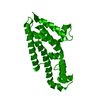

 PDBj
PDBj
 Assembly

 Mass: 58.693 Da / Num. of mol.: 3 / Source method: obtained synthetically / Formula: Ni
 Mass: 18.015 Da / Num. of mol.: 172 / Source method: isolated from a natural source / Formula: H2O
 Sample preparation
 / Beamline: ID14-2 / Wavelength: 0.933
  Processing